. 2022 Jul 5;23(13):7452.
doi: 10.3390/ijms23137452.

Lysophosphatidic Acid Is a Proinflammatory Stimulus of Renal Tubular Epithelial Cells

Affiliations

Lysophosphatidic Acid Is a Proinflammatory Stimulus of Renal Tubular Epithelial Cells

Christiana Magkrioti et al. Int J Mol Sci. .

Abstract

Chronic kidney disease (CKD) refers to a spectrum of diseases defined by renal fibrosis, permanent alterations in kidney structure, and low glomerular-filtration rate. Prolonged epithelial-tubular damage involves a series of changes that eventually lead to CKD, highlighting the importance of tubular epithelial cells in this process. Lysophosphatidic acid (LPA) is a bioactive lipid that signals mainly through its six cognate LPA receptors and is implicated in several chronic inflammatory pathological conditions. In this report, we have stimulated human proximal tubular epithelial cells (HKC-8) with LPA and 175 other possibly pathological stimuli, and simultaneously detected the levels of 27 intracellular phosphoproteins and 32 extracellular secreted molecules with multiplex ELISA. This quantification revealed a large amount of information concerning the signaling and the physiology of HKC-8 cells that can be extrapolated to other proximal tubular epithelial cells. LPA responses clustered with pro-inflammatory stimuli such as TNF and IL-1, promoting the phosphorylation of important inflammatory signaling hubs, including CREB1, ERK1, JUN, IκΒα, and MEK1, as well as the secretion of inflammatory factors of clinical relevance, including CCL2, CCL3, CXCL10, ICAM1, IL-6, and IL-8, most of them shown for the first time in proximal tubular epithelial cells. The identified LPA-induced signal-transduction pathways, which were pharmacologically validated, and the secretion of the inflammatory factors offer novel insights into the possible role of LPA in CKD pathogenesis.

Keywords: cytokines; inflammation; lysophosphatidic acid; tubular epithelial cells.


Conflict of interest statement

C.U. is an employee of F. Hoffmann-La Roche AG.

Figures

Figure 1
Figure 1
Multiplex ELISA for the detection of secreted factors and signaling molecules phosphorylation. Human kidney proximal tubular epithelial cells, HKC-8, were stimulated with 176 stimuli. Supernatants were collected at 24 h and cell lysates at 5 and 25 min post stimulation. Supernatants or cell lysates were added to a mix containing magnetic beads internally dyed with precise proportions of red and infrared fluorophores, thus, rendering unique spectral signature microspheres. Each unique microsphere-bead was conjugated with a distinct monoclonal antibody against a secreted factor or a phosphoprotein. Biotinylated detection antibodies were added to the mix, followed by a streptavidin-R-Phycoerythrin complex. This process allows the simultaneous recognition of 32 secreted factors or 27 phosphoproteins in one sample. Created with BioRender.com, accessed on 1 June 2022.
Figure 2
Figure 2
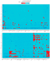
Differential expression of 32 secreted biological factors in the supernatants of human kidney proximal tubular epithelial cells (HKC-8) upon the stimulation with LPA (18:1) and 175 disparate biological stimuli. The expression was assessed with multiplex ELISA employing microbeads of unique spectral signatures conjugated with monoclonal antibodies specific for each of the 32 secreted factors. Red indicates active signals (FC ≥ 1.5). See also Figure S1.
Figure 3
Figure 3
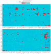
Phosphorylation of 27 major hubs in intracellular signaling pathways of human kidney proximal tubular epithelial cells (HKC-8) upon the stimulation with LPA (18:1) and 175 disparate biological stimuli. The expression was assessed with multiplex ELISA employing microbeads of unique spectral signatures conjugated with monoclonal antibodies specific for each of the 27 phosphoproteins. Red indicates active signals (FC ≥ 1.5). See also Figure S1.
Figure 4
Figure 4
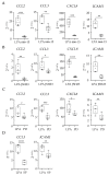
LPA stimulates the secretion of CCL2, CCL3, CXCL10, ICAM1, IL-6, and IL-8 from human kidney proximal tubular epithelial cells (HKC-8). Multiplex ELISA quantifying the expression of the indicated secreted factors in the supernatants from HKC-8 cells upon the stimulation with three different LPA species (16:0, 20:4, 18:1) at 10 μM for 24 h. Statistical significance was assessed with Brown–Forsythe’s and Welch’s ANOVA followed by Dunnett’s post hoc test in the case of normal distribution or with Kruskal–Wallis test in the case of non-normal distribution; * p < 0.05, ** p < 0.01, *** p < 0.001. Circles correspond to control values, squares correspond to LPA 16:0 values, upward triangles correspond to LPA 20:4 values and downward triangles correspond to LPA 18:1 values.
Figure 5
Figure 5
LPA stimulates the phosphorylation of JUN, IκΒα, MEK1, ERK1, and CREB1 in human kidney proximal tubular epithelial cells (HKC-8). Cells were incubated with three different LPA species (16:0, 20:4, 18:1) at 10 μΜ for 5 (A) or 25 min (B), and the phosphorylation was assessed with multiplex ELISA in triplicates. Circles correspond to control values, squares correspond to LPA 16:0 values, upward triangles correspond to LPA 20:4 values and downward triangles correspond to LPA 18:1 values. Statistical significance was assessed with Brown–Forsythe’s and Welch’s ANOVA followed by Dunnett’s post hoc test in the case of normal distribution or with Kruskal–Wallis test in the case of non-normal distribution. * p < 0.05, ** p < 0.01.
Figure 6
Figure 6
LPA clusters with proinflammatory stimuli. Heatmap of the active stimuli clustered in three major groups (1-3). Inactive stimuli and globally unresponsive signals were removed. Pairwise stimuli distance was calculated on binary transformed fold change values using Gower’s metric prior to divisive clustering. See also Figures S2–S4.
Figure 7
Figure 7
LPA stimulates the expression of CCL2, CCL3, CXCL8, CXCL10, ICAM1, and IL6 from human kidney proximal tubular epithelial cells (HKC-8). (A,B) HKC-8 cells were incubated for 1, 4, 12, and 24 h with 10 μM of LPA (A), and with 2.5, 5, and 10 μM LPA for 4 h (B). Control cells were stimulated with the equivalent volume of chloroform (VHC). mRNA-expression levels of the indicated secreted factors were quantified with RT-qPCR. The Cq values of each gene were normalized against the Cq values of B2M. The results represent the findings of two (A) and three (B) separate experiments. In (A) circles, upward triangles, downward triangles and diamonds refer to 1, 4, 12 and 24 hours of incubation with LPA, respectively. In (B) circles, upward triangles, downward triangles and diamonds refer to incubation with 0, 2.5, 5 and 10 μM LPA, respectively. Statistical significance was assessed in (A) with 2-way ANOVA and Tukey’s post hoc test and in (B) with Brown-Forsythe’s and Welch’s test or the Kruskal–Wallis test depending on the normality status of the data; * p < 0.05, ** p < 0.01, *** p < 0.001, **** p < 0.0001. See also Figure S5.
Figure 8
Figure 8
Pharmacologic dissection of LPA-induced cellular signaling pathways. HKC-8 cells were pretreated for 1 h with 666-15 (CREB1 inhibitor) 10 μM in (A), JSH23 (NFκΒ inhibitor) 100 μM in (B), PD98059 (MEK/ERK inhibitor) 50 μM in (C), or SP600125 (JNK inhibitor) 50 μM in (D) and then activated with LPA at a final concentration of 10 μΜ for 4 h. mRNA-expression levels of the indicated secreted factors were quantified with RT-qPCR. The Cq values of each gene were normalized against the Cq values of B2M. Statistical analysis was performed with unpaired t-test or Welch’s test in the case of normal data and with Mann–Whitney in the case of non-normal data. * p < 0.05, ** p < 0.01, *** p < 0.001, **** p < 0.0001. See also Figure S6.
Figure 9
Figure 9
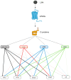
Graphical depiction of the LPA-induced signaling pathways in HKC-8 cells. LPA binds to the LPARs, which activate G proteins and the signal progresses to secondary signaling hubs, such as MEK/ERK or transcription factors c-JUN, CREB1, and NFκB. MEK/ERK, CREB1, and NFκB co-activate CCL2, CCL3, CXCL8 (IL-8), and ICAM1 expression. C-JUN activates only CCL3 and ICAM1 expression. Solid colored lines show connections that are derived from our results. Connections depicted with dashed lines are drawn from the literature and are not verified from our data. Created with BioRender.com, accessed on 1 June 2022.

References

    1. Lv J.C., Zhang L.X. Prevalence and Disease Burden of Chronic Kidney Disease. Adv. Exp. Med. Biol. 2019;1165:3–15. doi: 10.1007/978-981-13-8871-2_1. - DOI - PubMed
    1. Romagnani P., Remuzzi G., Glassock R., Levin A., Jager K.J., Tonelli M., Massy Z., Wanner C., Anders H.J. Chronic kidney disease. Nat. Rev. Dis. Primers. 2017;3:17088. doi: 10.1038/nrdp.2017.88. - DOI - PubMed
    1. Ruiz-Ortega M., Rayego-Mateos S., Lamas S., Ortiz A., Rodrigues-Diez R.R. Targeting the progression of chronic kidney disease. Nat. Rev. Nephrol. 2020;16:269–288. doi: 10.1038/s41581-019-0248-y. - DOI - PubMed
    1. Takaori K., Nakamura J., Yamamoto S., Nakata H., Sato Y., Takase M., Nameta M., Yamamoto T., Economides A.N., Kohno K., et al. Severity and Frequency of Proximal Tubule Injury Determines Renal Prognosis. J. Am. Soc. Nephrol. 2016;27:2393–2406. doi: 10.1681/ASN.2015060647. - DOI - PMC - PubMed
    1. Ferenbach D.A., Bonventre J.V. Mechanisms of maladaptive repair after AKI leading to accelerated kidney ageing and CKD. Nat. Rev. Nephrol. 2015;11:264–276. doi: 10.1038/nrneph.2015.3. - DOI - PMC - PubMed

MeSH terms

Substances