. 2022 Jun 24;15(13):4468.
doi: 10.3390/ma15134468.

Assessment of Angiogenesis and Cell Survivability of an Inkjet Bioprinted Biological Implant in an Animal Model

Affiliations

Assessment of Angiogenesis and Cell Survivability of an Inkjet Bioprinted Biological Implant in an Animal Model

Beu P Oropeza et al. Materials (Basel). .

Abstract

The rapidly growing field of tissue engineering hopes to soon address the shortage of transplantable tissues, allowing for precise control and fabrication that could be made for each specific patient. The protocols currently in place to print large-scale tissues have yet to address the main challenge of nutritional deficiencies in the central areas of the engineered tissue, causing necrosis deep within and rendering it ineffective. Bioprinted microvasculature has been proposed to encourage angiogenesis and facilitate the mobility of oxygen and nutrients throughout the engineered tissue. An implant made via an inkjet printing process containing human microvascular endothelial cells was placed in both B17-SCID and NSG-SGM3 animal models to determine the rate of angiogenesis and degree of cell survival. The implantable tissues were made using a combination of alginate and gelatin type B; all implants were printed via previously published procedures using a modified HP inkjet printer. Histopathological results show a dramatic increase in the average microvasculature formation for mice that received the printed constructs within the implant area when compared to the manual and control implants, indicating inkjet bioprinting technology can be effectively used for vascularization of engineered tissues.

Keywords: angiogenesis; bio-printing; inkjet printing; microvasculature; tissue engineering.


Conflict of interest statement

The authors declare no conflict of interest. The funders had no role in the design of the study; in the collection, analyses, or interpretation of data; in the writing of the manuscript, or in the decision to publish the results.

Figures

Figure 1
Figure 1
Schematic representation of implant placement in the in vivo model. The implant was inserted in a subcutaneous pocket in the dorsal thoracic area. The placement ensured the animal was not able to easily access the wound and cause harm to the area that was being monitored.
Figure 2
Figure 2
(A) In vivo tissue sections of the B17-SCID mice. Hematoxylin, eosin, and immunohistochemistry staining (blue-DAPI, green-CD31) visualize engineered vessels. Arrows indicate microvascular formations. (B) Blood vessel quantification of TIB implants show a 1.4× increase in the number of blood vessels per area compared to the manually made mock implants. The inkjet-printed implants presented 1.77× more vessels than printed mock implants. (C) Particle analysis of CD31-stained tissue samples show a 14× and 15× increase for the manually seeded and TIB samples, respectively. * denotes statistical significance p < 0.05.
Figure 3
Figure 3
(A) In vivo vascular formation for the NSG-SMG3 mice. Hematoxylin, eosin, and immunohistochemistry-stained (blue-DAPI, green-CD31). Arrows indicate microvascular formation. (B) For blood vessel quantification of NSG-SGM3 mice, we observed more than twice the amount of vessels when comparing the inkjet-printed implants to those from the mock control group; specifically, a 2.16 increase in vessels per area was counted. Similarly, 2.89× of vessels were seen for TIB implants vs. manually seeded implants in the NSG-SGM3. (C) Particle analyses of the CD31-stained NSG-SMG3 tissue sections display a 7.8× increase for the manually seeded cells when compared to the control group and a 40× increase when compared to the TIB group. * denotes statistical significance p < 0.05.
Figure 4
Figure 4
A comparison of the average number of vasculatures (A) and particle count for the CD31 stained sections (B) seen throughout the printed and manually seeded tissue sections for both types of mice (* p < 0.05, n = 3). All numbers were observed in 120 mm2 tissue sections.
Figure 5
Figure 5
(A) Images of inkjet printer cartridge nozzles under a light microscope. (B) Schematic representation of cells being printed through an inkjet nozzle.
Figure 6
Figure 6
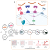
Angiogenesis development conceptual model. (A) Experimental procedure followed throughout the project, solid black arrows represent the observable progression of the project; dashed orange arrows denote a theoretical representation of angiogenic development. (B) Cellular pathway activation by thermal inkjet printing technologies, droplets of bioink are heated, binding VEGF-A to VEGFR, HSP27 to TLR3, HSP60 to TLR4, and HSP70 to TLR2/4, all leading to the angiogenesis seen in the analysis of tissues procured [20]. (C) Alginate/gelatin hydrogel degradation and vessel formation of the human microvascular endothelial cells via the inkjet-printing process.

References

    1. Mota C., Puppi D., Chiellini F., Chiellini E. Additive manufacturing techniques for the production of tissue engineering constructs. J. Tissue Eng. Regen. Med. 2015;9:174–190. doi: 10.1002/term.1635. - DOI - PubMed
    1. Matai I., Kaur G., Seyedsalehi A., McClinton A., Laurencin C.T. Progress in 3D bioprinting technology for tissue/organ regenerative engineering. Biomaterials. 2020;226:119536. doi: 10.1016/j.biomaterials.2019.119536. - DOI - PubMed
    1. Vacanti J.P., Morse M.A., Saltzman W.M., Domb A.J., Perez-Atayde A., Langer R. Selective cell transplantation using bioabsorbable artificial polymers as matrices. J. Pediatr. Surg. 1988;23:3–9. doi: 10.1016/S0022-3468(88)80529-3. - DOI - PubMed
    1. Mironov V., Kasyanov V., Drake C., Markwald R.R. Organ printing: Promises and challenges. Regen. Med. 2008;3:93–103. doi: 10.2217/17460751.3.1.93. - DOI - PubMed
    1. Richards D., Jia J., Yost M., Markwald R.R., Mei Y. 3D bioprinting for vascularized tissue fabrication. Ann. Biomed. Eng. 2017;45:132–147. doi: 10.1007/s10439-016-1653-z. - DOI - PMC - PubMed