. 2022 Jul 1;15(13):4655.
doi: 10.3390/ma15134655.

Biocompatibility of ZrO2 vs. Y-TZP Alloys: Influence of Their Composition and Surface Topography

Affiliations

Biocompatibility of ZrO2 vs. Y-TZP Alloys: Influence of Their Composition and Surface Topography

Alex Tchinda et al. Materials (Basel). .

Abstract

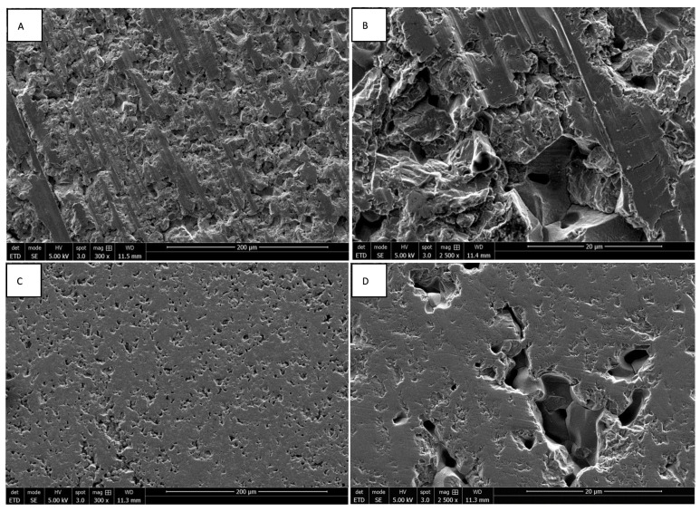
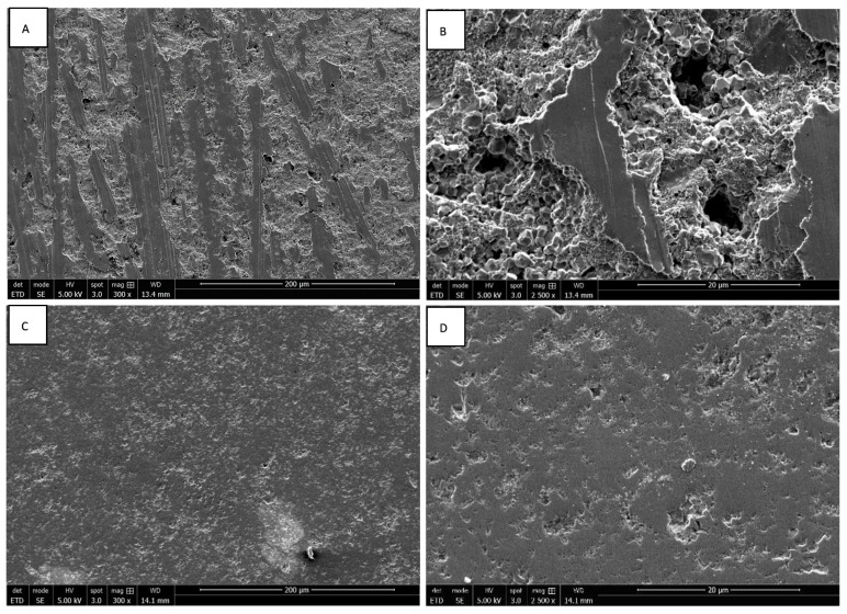
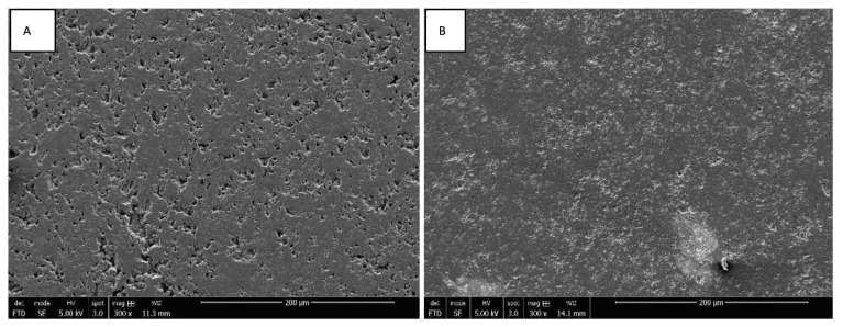
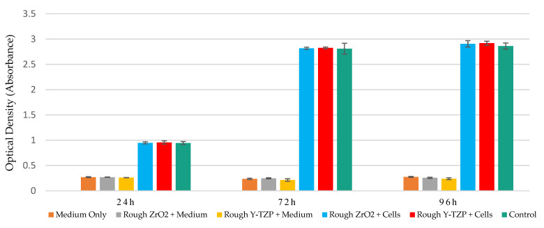
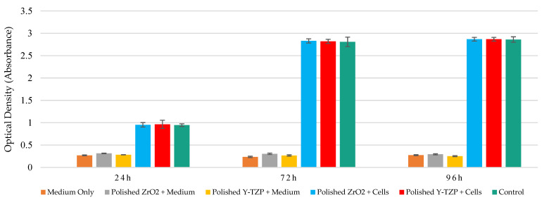
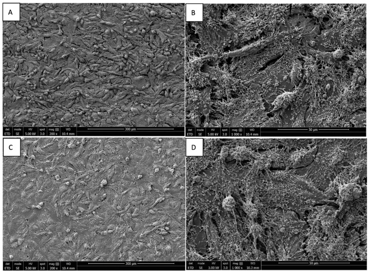
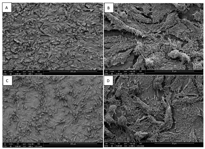
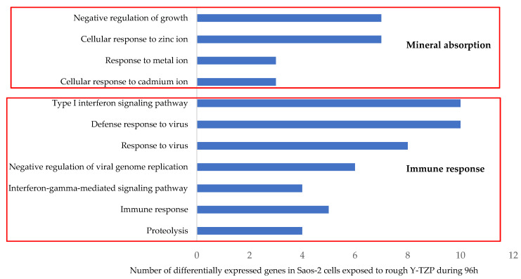
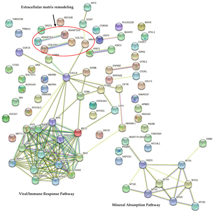
The osseointegration of implants is defined as the direct anatomical and functional connection between neoformed living bone and the surface of a supporting implant. The biological compatibility of implants depends on various parameters, such as the nature of the material, chemical composition, surface topography, chemistry and loading, surface treatment, and physical and mechanical properties. In this context, the objective of this study is to evaluate the biocompatibility of rough (Ra = 1 µm) and smooth (Ra = 0 µm) surface conditions of yttria-zirconia (Y-TZP) discs compared to pure zirconia (ZrO2) discs by combining a classical toxicological test, morphological observations by SEM, and a transcriptomic analysis on an in vitro model of human Saos-2 bone cells. Similar cell proliferation rates were observed between ZrO2 and Y-TZP discs and control cells, regardless of the surface topography, at up to 96 h of exposure. Dense cell matting was similarly observed on the surfaces of both materials. Interestingly, only 110 transcripts were differentially expressed across the human transcriptome, consistent with the excellent biocompatibility of Y-TZP reported in the literature. These deregulated transcripts are mainly involved in two pathways, the first being related to "mineral uptake" and the second being the "immune response". These observations suggest that Y-TZP is an interesting candidate for application in implantology.

Keywords: biocompatibility; morphology; osseointegration; proliferation; surface topography; transcriptome; yttria–zirconia; zirconia.


Conflict of interest statement

The authors declare no conflict of interest.

Figures

Figure 1
Figure 1
SEM images of rough ((A) magnification 300×; (B) magnification 2500×) and smooth surfaces ((C) magnification 300×; (D) magnification 2500×) of ZrO2 discs.
Figure 2
Figure 2
SEM images of rough ((A) magnification 300×; (B) magnification 2500×) and smooth surfaces ((C) magnification 300×; (D) magnification 2500×) of Y-TZP discs.
Figure 3
Figure 3
SEM images of smooth surfaces of ZrO2 ((A) magnification 300×) and Y-TZP ((B) magnification 300×) discs.
Figure 4
Figure 4
Metabolic activity monitoring (WST1 assay) of Saos-2 cells exposed to rough surfaces (Ra = 1 µm) of ZrO2 and Y-TZP for 24 h, 72 h, and 96 h (N = 4). ANOVA: Analysis of one-factor variances (ZrO2 vs. Y-TZP vs. controls). Not Significant at 24 h, 48 h, and 96 h.
Figure 5
Figure 5
Metabolic activity monitoring (WST1 assay) of Saos-2 cells exposed to mirror-polished surfaces (Ra = 0.01 µm) of ZrO2 and Y-TZP for 24 h, 72 h, and 96 h (N = 4). ANOVA: Analysis of one-factor variances (ZrO2 vs. Y-TZP vs. controls). Not Significant at 24 h, 72 h, or 96 h.
Figure 6
Figure 6
SEM image of Saos-2 cells on rough ((A) magnification 200×; (B) magnification 1000×) and mirror-polished ((C) magnification 200×; (D) magnification 1000×) surfaces of ZrO2 discs at 96 h of incubation.
Figure 7
Figure 7
SEM image of Saos-2 cells on rough ((A) magnification 200×; (B) magnification 1000×) and mirror-polished ((C) magnification 200×; (D) magnification 1000×) surfaces of Y-TZP discs at 96 h of incubation.
Figure 8
Figure 8
Enriched Gene Ontology Biological Processes (GO BPs) modified in Saos-2 cells exposed to rough surfaces (Ra = 1 µm) of Y-TZP discs for 96 h (determined through DAVID Bioinformatics Resources v6.8). (Z score > 1.3).
Figure 9
Figure 9
The STRING network of gene–gene interactions in Saos-2 cells exposed to the rough surfaces (Ra = 1 µm) of Y-TZP discs for 96 h (STRING database version 11.5). A list of all differentially expressed genes in Saos-2 cells exposed to the rough surfaces of Y-TZP discs for 96 h (FC ≥ |1.5, |f-value ≤ |0.01) (Supplementary Table S1) was injected into STRING database v11.5. The 2 significantly enriched KEGG pathways (determined using the DAVID database) are represented in bold. Legend: Network nodes represent proteins, edges represent protein–protein associations (red line: the presence of fusion evidence; green line: neighborhood evidence; blue line: co-occurrence evidence; purple line: experimental evidence; yellow line: text mining evidence; light blue line: database evidence; black line: co-expression evidence).

References

    1. Del Suárez-López Amo F., Garaicoa-Pazmiño C., Fretwurst T., Castilho R.M., Squarize C.H. Dental Implants-Associated Release of Titanium Particles: A Systematic Review. Clin. Oral Implant. Researc. 2018;29:1085–1100. doi: 10.1111/clr.13372. - DOI - PubMed
    1. Dias Corpa Tardelli J., da Lima Costa Valente M., de Theodoro Oliveira T., Cândido dos Reis A. Influence of Chemical Composition on Cell Viability on Titanium Surfaces: A Systematic Review. J. Prosthet. Dent. 2021;125:421–425. doi: 10.1016/j.prosdent.2020.02.001. - DOI - PubMed
    1. Jung R.E., Zembic A., Pjetursson B.E., Zwahlen M., Thoma D.S. Systematic Review of the Survival Rate and the Incidence of Biological, Technical, and Aesthetic Complications of Single Crowns on Implants Reported in Longitudinal Studies with a Mean Follow-up of 5 Years. Clin. Oral Implant. Res. 2012;23:2–21. doi: 10.1111/j.1600-0501.2012.02547.x. - DOI - PubMed
    1. Quentin G. Influence des Etats de Surface sur le Developpement des Peri Implantites. Volume 74. Université Claude Bernard-Lyon 1 U.F.R. d’odontologie; Lyon, France: 2014. [(accessed on 27 April 2022)]. Available online: http://bibnum.univ-lyon1.fr/nuxeo/nxfile/default/9a2e93b1-b59e-4efe-ae4a....
    1. Kissa J., El Kholti W., Chemlali S., Kawtari H., Laalou Y., Albandar J.M. Prevalence and Risk Indicators of Peri-implant Diseases in a Group of Moroccan Patients. J. Periodontol. 2021;92:1096–1106. doi: 10.1002/JPER.20-0549. - DOI - PubMed

LinkOut - more resources