. 2022 Jun 27;11(13):3722.
doi: 10.3390/jcm11133722.

Immunophenotypic Characteristics of Bone Marrow Microenvironment Cellular Composition at the Biochemical Progression of Multiple Myeloma

Affiliations

Immunophenotypic Characteristics of Bone Marrow Microenvironment Cellular Composition at the Biochemical Progression of Multiple Myeloma

Agnieszka Krzywdzińska et al. J Clin Med. .

Abstract

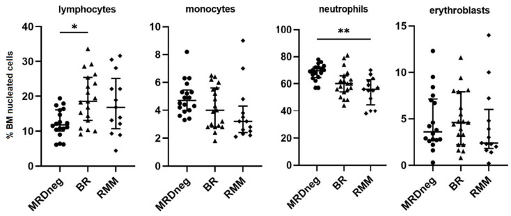
Multiple myeloma (MM) relapses are inevitable in the majority of patients, and in addition to genetic changes in the MM clone, the immune profile of the bone marrow (BM) plays a key role in this process. Biochemical progression or relapse (BR) precedes clinical relapse in a significant proportion of patients with MM. In the present study, we used flow cytometry to assess the cellular composition of the BM microenvironment in MM patients with confirmed BR. Fifteen distinct cells subsets in the BM were evaluated with the panel of antibodies used routinely for MRD monitoring in MM in 52 patients with MM (MRD-negative n = 20, BR n = 20, and clinically relapsed MM, RMM n = 12). The median percentage of MM cells detected in BR patients was 0.90% versus not detectable in MRD-negative patients and of 3.0% in RMM cohort. Compared to the MRD-negative group, BR status was associated with an increase in the percentage of lymphoid subpopulations, including memory B cells (p = 0.003), CD27+T cells (p = 0.002), and NK/NKT cells (p < 0.001). Moreover, a decrease in B-cell precursors (p < 0.001) and neutrophils (p = 0.006) was observed. There were no significant differences in the composition of the BM cell subpopulations between the BR and RMM groups. Our results indicate the involvement of B-, T-, and NK cells in the process of losing immune surveillance over the MM clone that leads to relapse. It can be speculated that similar studies of a larger cohort of BR patients can potentially identify a group of patients for which an early treatment intervention would be beneficial.

Keywords: biochemical relapse; immune profiling; microenvironment; multiple myeloma.


Conflict of interest statement

The authors declare no conflict of interest.

Figures

Figure 1
Figure 1
Gating strategy used for identification of the different populations of nuclear cells in BM from a representative patient in biochemical relapse of MM. (A) Determination of nucleated cells population by excluding doublets on FSC−H/FSC−A dot plot—gate singlets. (B) Total PCs population was determined by gating events CD38+highCD138+. (C) MM PCs (in red) were distinguished from normal PCs (in green) by aberrant immunophenotype: CD19−CD56+bright. (D) On the dot plot showing nuclear cells without the PCs population, B−cells region CD19 + SSClow was drawn. (E) On bivariate plot CD45 versus CD38 gated on B−cells regions defining mature B cells and B cell precursors were drawn. In the next graph (F) showing only mature B−cells, naive and memory B−cells were distinguished based on CD27 and CD81 expression. On bivariate plots gated on singlets and not PCs, myeloid precursors CD38+CD117+ (G) and mast cells CD117+bright (H) were identified. Among nucleated cells on the bivariate plot of CD45 versus SSC−A (I), population of erythroid CD45−/dim, polymorphonuclear cells (PMN), and mononuclear cells (MNC) were gated. On plot CD38 versus CD117, gated on erythroid cells erythroblasts and erythroid precursors CD117+ were identified (J). Population of PMN was divided on neutrophils and eosinophils based on the expression of CD81 and CD27 (K). In population of MNC, the lymphoid cells that express CD56 antigen were identified as NK/NKT cells (L). The rest of events without CD19 and CD56 expression and with lymphoid characteristic on CD45 versus SSC−A dot plot were classified as T cells (M). Next, a subpopulation of T cells with positive expression of CD27 was gated (N). Finally, on the bivariate plot of CD38 versus CD81, population of monocytes was precisely distinguished among CD45+SSCint nuclear cells (O). BM, bone marrow; MM, multiple myeloma; PCs, plasma cells.
Figure 2
Figure 2
Distribution of clonal plasma cells and normal plasma cells in BM samples from MRD negative patients in biochemical relapse (BR) and clinical relapsed MM (RMM). Graphs show the median and quartile Q1–Q3 values (** p < 0.01; *** p < 0.001). BM, bone marrow; MRD, measurable residual disease.
Figure 3
Figure 3
Correlation between percentage of MM plasma cells and the percentage of neutrophils, B−cell precursors, memory B cells, CD27+ T cells, and NK/NKT cells in bone marrow aspirates from MM patients: MRD negative (n = 20) with biochemical relapse (n = 20) and with clinical relapse (n = 12).
Figure 4
Figure 4
The differences in the median proportion of main BM populations: lymphocytes, monocytes, neutrophils, and erythroblasts between MRD negative patients with biochemical relapse (BR) and relapsed multiple myeloma (RMM) patients. Graphs show the median and quartile values Q1–Q3 (* p < 0.05; ** p < 0.01). BM, bone marrow; MRD, measurable residual disease.
Figure 5
Figure 5
The differences in the median proportion of BM lymphocytes subsets: B−cells precursors, naïve and memory B cells, CD27+T cells, and NK/NKT cells in MRD negative patients with biochemical relapse (BR) and relapsed multiple myeloma (RMM) patients. Graphs show the median and quartile Q1–Q3 values. (** p < 0.01; *** p < 0.001). BM, bone marrow; MRD, measurable residual disease.

Similar articles

Cited by

References

    1. Palumbo A., Anderson K. Multiple myeloma. N. Engl. J. Med. 2011;364:1046–1060. doi: 10.1056/NEJMra1011442. - DOI - PubMed
    1. Leone P., Solimando A.G., Malerba E., Fasano R., Buonavoglia A., Pappagallo F., De Re V., Argentiero A., Silvestris N., Vacca A., et al. Actors on the Scene: Immune cells in the myeloma niche. Front. Oncol. 2020;10:599098. doi: 10.3389/fonc.2020.599098. - DOI - PMC - PubMed
    1. Lopes R., Ferreira B.V., Caetano J., Barahona F., Carneiro E.A., João C. Boosting immunity against multiple myeloma. Cancers. 2021;13:1221. doi: 10.3390/cancers13061221. - DOI - PMC - PubMed
    1. Flores-Montero J., Sanoja-Flores L., Paiva B., Puig N., García-Sánchez O., Böttcher S., van der Velden V.H.J., Pérez-Morán J.J., Vidriales M.B., García-Sanz R., et al. Next generation flow for highly sensitive and standardized detection of minimal residual disease in multiple myeloma. Leukemia. 2017;31:2094–2103. doi: 10.1038/leu.2017.29. - DOI - PMC - PubMed
    1. Munshi N.C., Avet-Loiseau H., Anderson K.C., Neri P., Paiva B., Samur M., Dimopoulos M., Kulakova M., Lam A., Hashim M., et al. Large meta-analysis establishes the role of MRD negativity in long-term survival outcomes in multiple myeloma patients. Blood Adv. 2020;4:5988–5999. doi: 10.1182/bloodadvances.2020002827. - DOI - PMC - PubMed