. 2022 Jun 21;27(13):3991.
doi: 10.3390/molecules27133991.

Computational Prediction and Experimental Validation of the Unique Molecular Mode of Action of Scoulerine

Affiliations

Computational Prediction and Experimental Validation of the Unique Molecular Mode of Action of Scoulerine

Mahshad Moshari et al. Molecules. .

Abstract

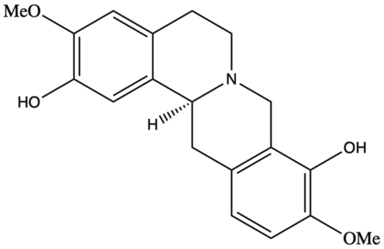
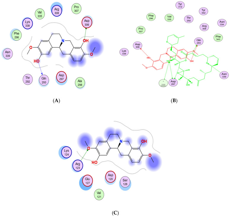
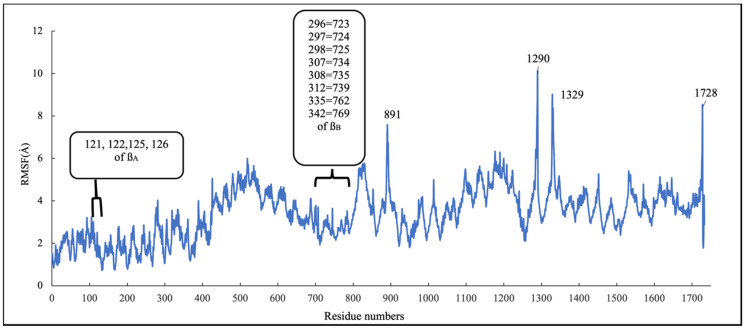
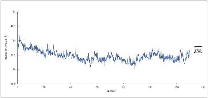
Scoulerine is a natural compound that is known to bind to tubulin and has anti-mitotic properties demonstrated in various cancer cells. Its molecular mode of action has not been precisely known. In this work, we perform computational prediction and experimental validation of the mode of action of scoulerine. Based on the existing data in the Protein Data Bank (PDB) and using homology modeling, we create human tubulin structures corresponding to both free tubulin dimers and tubulin in a microtubule. We then perform docking of the optimized structure of scoulerine and find the highest affinity binding sites located in both the free tubulin and in a microtubule. We conclude that binding in the vicinity of the colchicine binding site and near the laulimalide binding site are the most likely locations for scoulerine interacting with tubulin. Thermophoresis assays using scoulerine and tubulin in both free and polymerized form confirm these computational predictions. We conclude that scoulerine exhibits a unique property of a dual mode of action with both microtubule stabilization and tubulin polymerization inhibition, both of which have similar affinity values.

Keywords: cancer treatment; drug discovery; microtubule; molecular dynamic simulation; protein docking; scoulerine.


Conflict of interest statement

The authors declare no competing interests.

Figures

Figure 1
Figure 1
Scoulerine structure.
Figure 2
Figure 2
S1, S2, and S3 represent the three predicted potential binding sites by blind docking of scoulerine (blue) to α (green) and β (red) tubulins of 1SA0 PDB structure. Colchicine derivative from 1SA0 in S1 and laulimalide from 404H in S2 are shown in white.
Figure 3
Figure 3
Interactions between the pharmacophoric points and the tubulin structure. Seven pharmacophoric points: three hydrogen bond acceptors (A1, A2, and A3) in purple dots, one hydrogen bond donor (D1) in an orange dot, two hydrophobic centers (H1 and H2) in green dots, and one planar group (R1) in a red dot. Figure 3 was designed based on the information provided in the source [25].
Figure 4
Figure 4
(A) Two-dimensional interaction scheme of the superimposed colchicine crystal structure from the 5NM5 PDB file (green) on scoulerine in the S1 site (red) on 1SA0. Star (*) on residue 318 indicates two different amino acids on 5NM5 and 1SA0 structures. (B) Two-dimensional interaction scheme of scoulerine in the S1 site. (C) Surface patches identifying regions of hydrophobicity (yellow) around scoulerine. Residues Leu255, Ala316, Val318, and Ile378 of β tubulin that are involved in hydrophobic interactions are colored in teal.
Figure 5
Figure 5
RMSD of scoulerine in the colchicine binding site.
Figure 6
Figure 6
RMSF of all residues and scoulerine in the colchicine binding site.
Figure 7
Figure 7
The radius of gyration of all residues and scoulerine in the colchicine binding site.
Figure 8
Figure 8
Mass-weighted root–mean–squared deviation (Å) of the binding site of colchicine on tubulin, classified according to the cluster number, with occupancy indicated. The binding site includes scoulerine and residues having atoms within 8 Å of scoulerine. The dark blue part of the graph illustrates the equilibration phase of the simulation.
Figure 9
Figure 9
(A) Representative structures of scoulerine in cluster B (purple) versus colchicine (yellow). (B) Two-dimensional interaction scheme of scoulerine in the colchicine binding site. (C) Representative structures of cluster A (red), cluster B (purple), and cluster C (dark pink) in the colchicine binding site, α tubulin colored in teal, and βI tubulin colored in light pink. (D) Colchicine (yellow) in the colchicine binding site, α tubulin colored in teal, and βI tubulin colored in light pink. (E) Surface patches identifying regions of hydrophobicity (yellow) around scoulerine, residues Leu255, Val318, and Ile378 of β tubulin that are involved in hydrophobic interaction colored in teal.
Figure 10
Figure 10
Laulimalide in the laulimalide binding site of β tubulin (green) based on the 4O4H PDB file. The residues in blue are involved hydrogen bond interaction with laulimalide (purple).
Figure 11
Figure 11
(A) Two-dimensional interaction scheme of scoulerine in the S2 site identified by blind docking. (B) Two-dimensional interaction scheme of the superimposed laulimalide crystal structure based on the 4O4H PDB file (green) with scoulerine (red) in the S2 site on 1SA0. Star (*) on residue 298 indicates two different amino acids on 4O4H and 1SA0 structures. (C) Two-dimensional interaction scheme of scoulerine in the S3 site found via blind docking.
Figure 12
Figure 12
RMSD plot of scoulerine docked to the laulimalide binding sites.
Figure 13
Figure 13
RMSF of all residues and scoulerine in laulimalide binding sites.
Figure 14
Figure 14
The radius of gyration of all residues and scoulerine in the laulimalide binding sites.
Figure 15
Figure 15
Mass-weighted root–mean–squared deviation (Å) of the binding sites of laulimalide to tubulin, classified according to cluster number, with their occupancy indicated. The binding site includes scoulerine and tubulin residues whose atoms are within 8 Å of scoulerine. The purple part of the graph illustrates the equilibration phase of the simulation.
Figure 16
Figure 16
(A) Three-dimensional interaction scheme of scoulerine (blue) and a superimposed laulimalide crystal structure from the 4O4H PDB file (purple) between microtubule protofilaments. Residues shown in light green are in the laulimalide site on βA tubulin and residues shown in dark green are in the laulimalide site on βB tubulin. (B) Two-dimensional interaction scheme of scoulerine in the laulimalide binding sites on βA tubulin and βB tubulin. (C) Representative structures of cluster A (purple) and cluster B (pink) in the laulimalide binding sites. αA and αB tubulins colored in light and dark pink and βA and βB tubulins colored in light and dark green, respectively.
Figure 17
Figure 17
(A) Purified porcine αβ tubulin (colchicine binding site, blue circle) and microtubule (laulimalide binding site, red square) binding to scoulerine via microscale thermophoresis. Each data point represents the mean of two independent measurements and the error bar is shown as the standard deviation. The binding curve is fitted with Graphpad Prism 7.0. (B) Fluorescence labelled purified porcine α/β tubulin binding to colchicine. Normalized microscale thermophoresis time traces are shown on the right. Each data point is the mean of three independent microscale thermophoresis measurements; error bars represent the standard deviation. The binding curve is fitted with Graphpad Prism 7.0.

References

    1. Cragg G.M., Pezzuto J.M. Natural products as a vital source for the discovery of cancer chemotherapeutic and chemopreventive agents. Med. Princ. Pract. 2016;25:41–59. doi: 10.1159/000443404. - DOI - PMC - PubMed
    1. Liu D., Meng X., Wu D., Qiu Z., Luo H. A Natural isoquinoline alkaloid with antitumor activity: Studies of the biological activities of berberine. Front. Pharmacol. 2019;10:9. doi: 10.3389/fphar.2019.00009. - DOI - PMC - PubMed
    1. Tian J., Mo J., Xu L., Zhang R., Qiao Y., Liu B., Jiang L., Ma S., Shi G. Scoulerine promotes cell viability reduction and apoptosis by activating ROS-dependent endoplasmic reticulum stress in colorectal cancer cells. Chem. Biol. Interact. 2020;327:109184. doi: 10.1016/j.cbi.2020.109184. - DOI - PubMed
    1. Kukula-Koch W.A., Widelski J. Alkaloids. In: Badal S., Delgoda R., editors. Pharmacognosy. Elsevier; Boston, MA, USA: 2017. pp. 163–198.
    1. Habartova K., Havelek R., Seifrtova M., Kralovec K., Cahlikova L., Chlebek J., Cermakova E., Mazankova N., Marikova J., Kunes J., et al. Scoulerine affects microtubule structure, inhibits proliferation, arrests cell cycle and thus culminates in the apoptotic death of cancer cells. Sci. Rep. 2018;8:4829. doi: 10.1038/s41598-018-22862-0. - DOI - PMC - PubMed

LinkOut - more resources