. 2022 Jun 21;22(13):4678.
doi: 10.3390/s22134678.

Evaluation of a High-Sensitivity Organ-Targeted PET Camera

Affiliations

Evaluation of a High-Sensitivity Organ-Targeted PET Camera

Justin Stiles et al. Sensors (Basel). .

Abstract

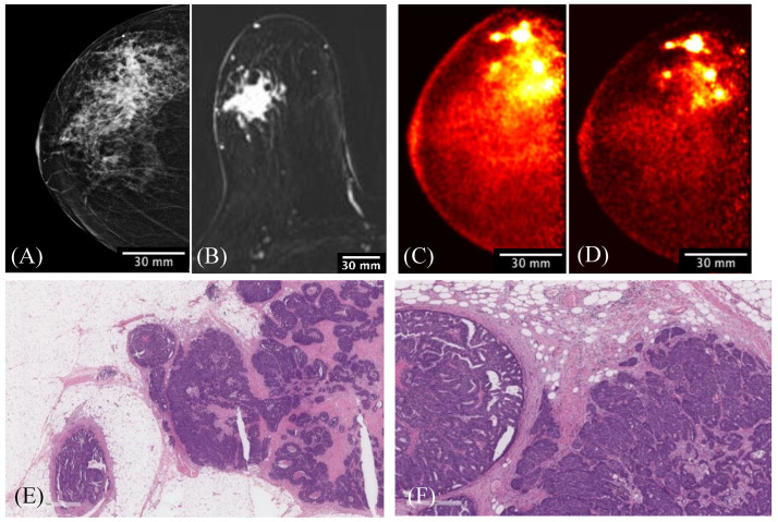
The aim of this study is to evaluate the performance of the Radialis organ-targeted positron emission tomography (PET) Camera with standardized tests and through assessment of clinical-imaging results. Sensitivity, count-rate performance, and spatial resolution were evaluated according to the National Electrical Manufacturers Association (NEMA) NU-4 standards, with necessary modifications to accommodate the planar detector design. The detectability of small objects was shown with micro hotspot phantom images. The clinical performance of the camera was also demonstrated through breast cancer images acquired with varying injected doses of 2-[fluorine-18]-fluoro-2-deoxy-D-glucose (18F-FDG) and qualitatively compared with sample digital full-field mammography, magnetic resonance imaging (MRI), and whole-body (WB) PET images. Micro hotspot phantom sources were visualized down to 1.35 mm-diameter rods. Spatial resolution was calculated to be 2.3 ± 0.1 mm for the in-plane resolution and 6.8 ± 0.1 mm for the cross-plane resolution using maximum likelihood expectation maximization (MLEM) reconstruction. The system peak noise equivalent count rate was 17.8 kcps at a 18F-FDG concentration of 10.5 kBq/mL. System scatter fraction was 24%. The overall efficiency at the peak noise equivalent count rate was 5400 cps/MBq. The maximum axial sensitivity achieved was 3.5%, with an average system sensitivity of 2.4%. Selected results from clinical trials demonstrate capability of imaging lesions at the chest wall and identifying false-negative X-ray findings and false-positive MRI findings, even at up to a 10-fold dose reduction in comparison with standard 18F-FDG doses (i.e., at 37 MBq or 1 mCi). The evaluation of the organ-targeted Radialis PET Camera indicates that it is a promising technology for high-image-quality, low-dose PET imaging. High-efficiency radiotracer detection also opens an opportunity to reduce administered doses of radiopharmaceuticals and, therefore, patient exposure to radiation.

Keywords: breast cancer; cancer detection; detectors; functional imaging; low-dose imaging; organ-targeted PET; precision medicine.


Conflict of interest statement

O.B., B.B., M.W., and A.R. have a financial interest in Radialis Medical. The funders had no role in the design of the study; in the collection, analyses, or interpretation of data; in the writing of the manuscript, or in the decision to publish the results.

Figures

Figure 1
Figure 1
Configuration of the Radialis PET Camera with two planar detector heads to be positioned on either side of a breast.
Figure 2
Figure 2
(A) Top view of 3 × 4 array of sensor modules inside a detector head; (B) photo of a block detector with the crystal array wrapped in a light-reflective material and an electronic board underneath.
Figure 3
Figure 3
Schematic representation of a cross-sectional view of three tiled block detectors.
Figure 4
Figure 4
Schematic representation of the overall detector field of view and axis convention.
Figure 5
Figure 5
System spatial resolutions produced with the MLEM reconstruction. Left: the central Z-axis resolution plotted as a function of point-source location along the X-axis. Right: the quarter Z-axis resolution plotted as a function of point-source location along the X-axis.
Figure 6
Figure 6
System spatial resolutions produced with a back-projection reconstruction. Left: the central Z-axis resolution plotted as a function of point-source location along the X-axis. Right: the quarter of the Z-axis resolution plotted as a function of point-source location along the X-axis.
Figure 7
Figure 7
Images of the micro hotspot phantom reconstructed using an MLEM reconstruction (left) and with a back-projection reconstruction (right).
Figure 8
Figure 8
Axial absolute sensitivity plotted against point-source location along the X-axis.
Figure 9
Figure 9
System performance count rates for a 90 mm LOR angle allowance. Left: low range of activity concentrations. Right: full range of activity concentrations.
Figure 10
Figure 10
A 56-year-old female with invasive ductal carcinoma and intermediate-grade DCIS. Digital mammography of right breast (A) and right breast Radialis PET image with 37 MBq 18F-FDG injection (B) both in the same projection (CC view) are presented for comparison between these two imaging modalities. Cancers are demonstrated by the arrows (A,B) and arrowhead (B). The second cancer (arrowhead) is visualized only by Radialis PET (B).
Figure 11
Figure 11
A 61-year-old female with right-breast multifocal invasive and in situ ductal carcinoma. Images of the same breasts in (A) FFDM in the CC plane showing extensive distortion, (B) a selected slice of MRI in the axial plane showing one irregular shape-enhancing mass lesion after 2 min post gadolinium-chelate-based contrast administration, (C) 3D Radialis PET in the CC plane where multiple distinct regions of contrast uptake after 1 h of 178 MBq 18F-FDG injection are evident, (D) 3D Radialis PET in the CC plane where the conspicuity of the multiple regions of enhanced 18F-FDG uptake (indicative of multifocal cancers) remains after 3 h from the prior (C) acquisition, (E) invasive carcinoma in the center of the field with in situ carcinoma present at the periphery in the pathology of the mastectomy specimen, and (F) higher-power view demonstrating intermediate-grade invasive carcinoma on the right and papillary ductal carcinoma in situ on the left.
Figure 12
Figure 12
The MLO-view digital mammography image (A) demonstrates the palpable mass (red circle) associated with the radiopaque marker placed on the patient’s skin. The presented slice of Radialis PET Camera CC image with 200 MBq injected 18F-FDG (B) identifies this lesion against the chest wall as well as two additional posterior masses. (C) View of the largest focus showing invasive ductal carcinoma of no special type; a clip-site reaction is present in the center of the tumor. (D) Second invasive focus demonstrating similar morphologic features and histologic grade. The 3 regions of contrast enhancement identified by Radialis PET are all biopsy-confirmed cancers.
Figure 13
Figure 13
A 33-year-old high-risk female underwent pre-operative breast MRI with multiplicity of enhancing masses demonstrated by the 3D-MIP image (A) and without corresponding masses demonstrated by the Radialis PET Camera images (B) with a 43 MBq injection. The mediolateral oblique views from the Radialis PET Camera are presented for the left (B) and right (C) side without evident focal 18F-FDG uptake in either image. The surgical pathology results do not show signs of cancer.
Figure 14
Figure 14
Side-by-side comparison of 307 MBq PET images from a breast cancer patient scanned with a Siemens Biograph PET/CT reconstructed using a time-of-flight reconstruction technique (TOF) (A,B) and with the Radialis PET system (C).

References

    1. Subramaniam R.M. Precision Medicine and PET/Computed Tomography: Challenges and Implementation. PET Clin. 2017;12:1–5. doi: 10.1016/j.cpet.2016.08.010. - DOI - PMC - PubMed
    1. Miles K.A., Voo S.A., Groves A.M. Additional Clinical Value for PET/MRI in Oncology: Moving Beyond Simple Diagnosis. J. Nucl. Med. 2018;59:1028–1032. doi: 10.2967/jnumed.117.203612. - DOI - PubMed
    1. Subramaniam R.M. Nuclear Medicine and Molecular Imaging—An Impactful Decade of Contributions to Patient Care and Driving Precision Medicine. Am. J. Roentgenol. 2017;209:241–242. doi: 10.2214/AJR.17.18569. - DOI - PubMed
    1. Wang X., Zhong X., Cheng L. Titanium-based nanomaterials for cancer theranostics. Coord. Chem. Rev. 2021;430:213662. doi: 10.1016/j.ccr.2020.213662. - DOI
    1. Miyake K.K., Nakamoto Y., Togashi K. Current Status of Dedicated Breast PET Imaging. Curr Radiol Rep. 2016;4:16. doi: 10.1007/s40134-016-0145-0. - DOI

Substances