

Form Factors as Potential Imaging Biomarkers to Differentiate Benign vs. Malignant Lung Lesions on CT Scans
- PMID: 35808538
- PMCID: PMC9269784
- DOI: 10.3390/s22135044
Form Factors as Potential Imaging Biomarkers to Differentiate Benign vs. Malignant Lung Lesions on CT Scans
Abstract
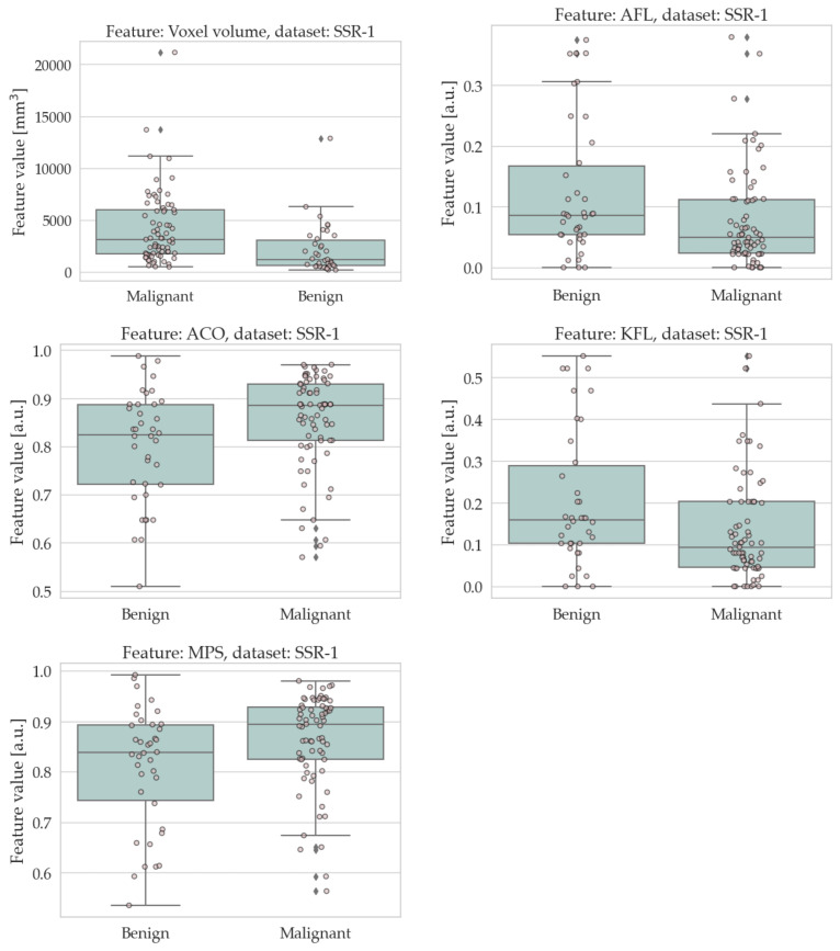
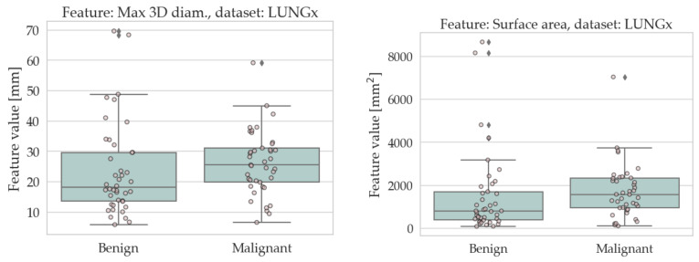
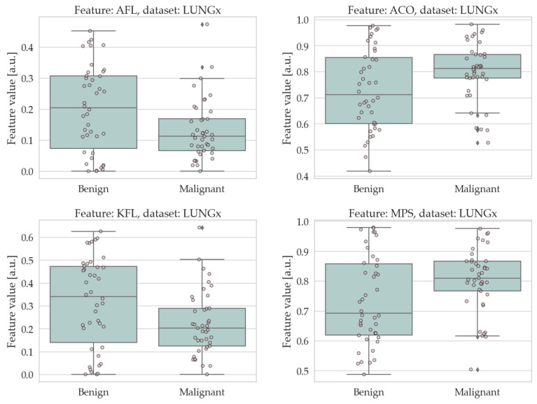
Indeterminate lung nodules detected on CT scans are common findings in clinical practice. Their correct assessment is critical, as early diagnosis of malignancy is crucial to maximise the treatment outcome. In this work, we evaluated the role of form factors as imaging biomarkers to differentiate benign vs. malignant lung lesions on CT scans. We tested a total of three conventional imaging features, six form factors, and two shape features for significant differences between benign and malignant lung lesions on CT scans. The study population consisted of 192 lung nodules from two independent datasets, containing 109 (38 benign, 71 malignant) and 83 (42 benign, 41 malignant) lung lesions, respectively. The standard of reference was either histological evaluation or stability on radiological followup. The statistical significance was determined via the Mann-Whitney U nonparametric test, and the ability of the form factors to discriminate a benign vs. a malignant lesion was assessed through multivariate prediction models based on Support Vector Machines. The univariate analysis returned four form factors (Angelidakis compactness and flatness, Kong flatness, and maximum projection sphericity) that were significantly different between the benign and malignant group in both datasets. In particular, we found that the benign lesions were on average flatter than the malignant ones; conversely, the malignant ones were on average more compact (isotropic) than the benign ones. The multivariate prediction models showed that adding form factors to conventional imaging features improved the prediction accuracy by up to 14.5 pp. We conclude that form factors evaluated on lung nodules on CT scans can improve the differential diagnosis between benign and malignant lesions.
Keywords: computed tomography; form factors; lung cancer; radiomics.
Conflict of interest statement
The authors declare no conflict of interest.
Figures
References
-
- World Health Organization Cancer. 2021. [(accessed on 24 August 2021)]. Available online: https://www.who.int/news-room/fact-sheets/detail/cancer.
-
- American Cancer Society Key Statistics for Lung Cancer. 2022. [(accessed on 24 June 2022)]. Available online: https://www.cancer.org/cancer/lung-cancer/about/key-statistics.html.
-
- Altavilla G., Di Maio M. I Numeri del Cancro in Italia. Intermedia Editore; Brescia, Italy: 2022. Polmone; pp. 56–57. Chapter 3.6.
MeSH terms
Substances
Grants and funding
LinkOut - more resources
Full Text Sources
Medical
Miscellaneous
