. 2022 Jul 1;14(13):2706.
doi: 10.3390/polym14132706.

Supercritical Impregnation of Mango Leaf Extract into PLA 3D-Printed Devices and Evaluation of Their Biocompatibility with Endothelial Cell Cultures

Affiliations

Supercritical Impregnation of Mango Leaf Extract into PLA 3D-Printed Devices and Evaluation of Their Biocompatibility with Endothelial Cell Cultures

Pilar Grosso et al. Polymers (Basel). .

Abstract

The addition of natural substances with pharmacoactive properties to polymeric biomedical devices would provide beneficial regarding the assimilation of these endoprostheses when implanted into a patient's body. The added drug would facilitate endothelization by regulating the inflammatory processes that such interventions entail, preventing contamination hazards and favoring the angiogenesis or formation of blood vessels in the tissue. The present work used mango leaf extract (MLE) obtained through pressurized ethanol for this purpose. Polylactic acid (PLA) in the form of filaments or 3D-printed disks was impregnated by means of supercritical technology with MLE for the culture essays. The release kinetics has been studied and the polymer matrices have been examined by scanning electron microscopy (SEM). The impregnated devices were subjected to in vitro culture of colony-forming endothelial cells. The influence of the different impregnation conditions used for the production of the MLE impregnated polymeric devices on the development of the cell culture was determined by fluorescence microscopy. The best results were obtained from the calcein cultures on 35 °C MLE impregnated into 3D-printed polymer disks.

Keywords: 3D printer; ECFCs; PLA; mango leaf; supercritical impregnation.


Conflict of interest statement

The authors declare no conflict of interest. The funders had no role in the design of the study, in the collection, analyses, or interpretation of the data, in the writing of the manuscript, or in the decision to publish the results.

Figures

Figure 1
Figure 1
Flow diagram of the supercritical extraction and impregnation equipment.
Figure 2
Figure 2
Polymer metal supports used over the impregnation process.
Figure 3
Figure 3
The 3D virtual image (a) and photographs (b) of freshly printed disks and after being used for the cell proliferation assays.
Figure 4
Figure 4
PLA filaments impregnated with MLE: (a) 35 °C, 100 bar; (b) 35 °C, 400 bar; (c) 55 °C, 400 bar; and (d) 55 °C, 100 bar.
Figure 5
Figure 5
Release kinetics of 3D + SCI samples produced under different pressure and temperature conditions.
Figure 6
Figure 6
Release kinetics of SCI + 3D samples produced under different pressure and temperature conditions.
Figure 7
Figure 7
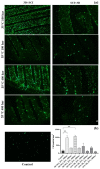
Viability of ECFCs grown on MLE impregnated PLA disks. (a) Representative images of ECFCs cultured on PLA disks impregnated with mango extracts under different sets of conditions, compared against a non-impregnated control disc. Calcein labeling can be observed (green dots) after incubation for 1 h at 37 °C. The photographs were taken at 4× magnification. (b) Graphical representation of the cell count per unit area under the different impregnating conditions of the PLA disks. The values are represented as mean ± SEM (n = 3). ** p-value < 0.01.
Figure 8
Figure 8
Analysis of the cell morphology of the ECFCs cultured on PLA disks impregnated with mango extract. Images of the ECFCs cultures on PLA disks impregnated with mango extracts under different conditions and non-impregnated control disc. The photographs were taken at 10× magnification: (a) PLA control; (b) 35 °C, 100 bar (3D + SCI); (c) 35 °C, 400 bar (3D + SCI); (d) 35 °C, 400 bar (SCI + 3D).
Figure 9
Figure 9
Scanning electron microscopy examination of 3D + SCI samples: (a) PLA control disks (100×, 400×, 800×); (b) 35 °C, 100 bar (100×, 400×, 400×); and (c) 35 °C, 400 bar (100×, 800×, 800×).

References

    1. Birajdar M.S., Joo H., Koh W.G., Park H. Natural bio-based monomers for biomedical applications: A review. Biomater. Res. 2021;25:8. doi: 10.1186/s40824-021-00208-8. - DOI - PMC - PubMed
    1. Ahmed K.K., Tamer M.A., Ghareeb M.M., Salem A.K. Recent Advances in Polymeric Implants. AAPS PharmSciTech. 2019;20:300. doi: 10.1208/s12249-019-1510-0. - DOI - PubMed
    1. Ventola C.L. Medical Applications for 3D Printing: Current and Projected Uses. Pharm. Ther. 2014;39:704–711. - PMC - PubMed
    1. Zhu X., Li H., Huang L., Zhang M., Fan W., Cui L. 3D printing promotes the development of drugs. Biomed. Pharmacother. 2020;131:110664. doi: 10.1016/j.biopha.2020.110644. - DOI - PubMed
    1. Nugroho W.T., Dong Y., Pramanik A. Dimensional accuracy and surface finish of 3D printed polyurethane (PU) dog-bone samples optimally manufactured by fused deposition modelling (FDM) Rapid Prototyp. J. 2022 doi: 10.1108/RPJ-12-2021-0328. in press . - DOI