Review
. 2022 Jul 5;14(13):2751.
doi: 10.3390/polym14132751.

An Overview of In Vitro Drug Release Methods for Drug-Eluting Stents

Affiliations
Review

An Overview of In Vitro Drug Release Methods for Drug-Eluting Stents

Navideh Abbasnezhad et al. Polymers (Basel). .

Abstract

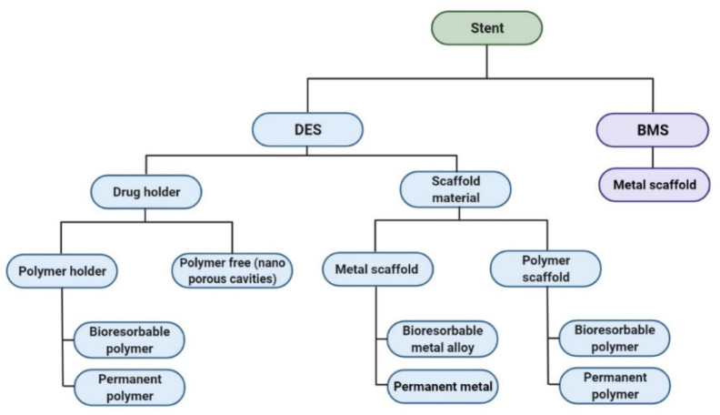
The drug release profile of drug-eluting stents (DESs) is affected by a number of factors, including the formulation, design, and physicochemical properties of the utilized material. DES has been around for twenty years and despite its widespread clinical use, and efficacy in lowering the rate of target lesion restenosis, it still requires additional development to reduce side effects and provide long-term clinical stability. Unfortunately, for analyzing these implants, there is still no globally accepted in vitro test method. This is owing to the stent's complexity as well as the dynamic arterial compartments of the blood and vascular wall. The former is the source of numerous biological, chemical, and physical mechanisms that are more commonly observed in tissue, lumen, and DES. As a result, universalizing bio-relevant apparatus, suitable for liberation testing of such complex implants is difficult. This article aims to provide a comprehensive review of the methods used for in vitro release testing of DESs. Aspects related to the correlation of the release profiles in the cases of in vitro and in vivo are also addressed.

Keywords: IVIV correlation; drug-eluting stents; flow conditions; hydrogels; in vitro release testing.


Conflict of interest statement

The authors declare that they have no conflict of interest.

Figures

Figure 1
Figure 1
Coronary restenosis after coronary angioplasty with the balloon, Reproduced with permission from [3].
Figure 2
Figure 2
In-stents coronary restenosis after coronary angioplasty with the bare metal stents, reproduced with permission from [3].
Figure 3
Figure 3
Late thrombosis after drug-eluting stents, reproduced with permission from and modified from [3].
Figure 4
Figure 4
Schematic of the material choice in the various stents [19,23,24,25,26,27,28,29,30].
Figure 5
Figure 5
Different geometries of stents (a) cylindrical (1. Glass beads, 2. acrylic glass disc, 3. hydrogel and 4. expanded stent) and (b) rectangular used in different studies, reproduced with permission from [46,47]. Reproduced with permission from Anne Seidlitz, Stefan Nagel, Beatrice Semmling, Niels Grabow, Heiner Martin, Volkmar Senz, Claus Harder, Katrin, Sternberg, Klaus-Peter Schmitz, Heyo K. Kroemer, Werner Weitschies, Examination of drug release and distribution from drug-elutingstents with a vessel-simulating flow-through cell; published by Elsevier, 2011.
Figure 6
Figure 6
Contribution of different mechanisms at different compartments, affecting the drug release from DES.
Figure 7
Figure 7
Photographs of freshly prepared native hydrogels of the final gel formulations: 3 wt.% alginate, 2 wt.% agar, 2 wt.% agarose, 10 wt.% PAA and 15 wt.% PVA (from left to right), reproduced with permission from [59].
Figure 8
Figure 8
Schematic drawing of the flow through cell: 1 large glass bead; 2 small glass beads; 3 metal disc; 4 hydrogel matrix; 5 drug eluting balloon (DEB), reprinted from [61].
Figure 9
Figure 9
Comparison of reference and modified hydrogels. Alginate gel versus LiChroprep gel containing 5% w/w LiChroprep® RP-18. Cumulative amounts (%) of triamterene detected in PBS pH 7.4, respective gel formulations, and residual model substance fractions within the stent coating, reprinted from [64].
Figure 10
Figure 10
(a) Experimental test bench, (b) pulsatile and steady inlet flow rate waveforms used in this study, reprinted from [42].
Figure 11
Figure 11
Schematic overview of the in vitro test setup (A) and photograph of the vFTC equipped with a 2 wt% agarose gel (B); (1) vFTC, (2) media container, (3) PBS of pH 7.4, (4) paddle stirred at 50 rpm, (5) peristaltic pump, (6) heated water bath, (a) glass beads, (b) stainless steel disc, (c) hydrogel, reproduced with permission from [59].
Figure 12
Figure 12
Normalized release of triamterene from stent coatings into media over time with vessel-simulating flow-through cell, flow-through cell (USP 4), or paddle apparatus (USP 2); flow rate 35 mL/min, paddle speed 50 rpm, reprinted from [85].
Figure 13
Figure 13
Illustration of convolution and deconvolution in IVIVC development, Reproduced with permission from [100]. Reproduced with permission from Y. Qiu, J.Z. Duan, Developing Solid Oral Dosage Forms; published by Elsevier, 2017.
Figure 14
Figure 14
(a) In vitro and (b) in vivo, release profiles of paclitaxel and sirolimus from PLGA/ACP coated stents, reprinted from [81].
Figure 15
Figure 15
Comparison of the in vitro and in vivo profiles with time-scaling factor, reprinted from [56].
Figure 16
Figure 16
Comparison between experimental in vitro drug release data (normalized by the cumulative mass eluted by the final measurement time point), and model simulations. LEFT: The low dose stent was well-fitted by a diffusion coefficient of the order of 10−16 m2s−1. RIGHT: The fit between the diffusion model and the data from the high dose stent was not so good. Furthermore, the best-fitting diffusion coefficient was of the order 10−17 m2s−1, reprinted from [108]. Reproduced with permission from Craig M. McKittrick, Sean McKee, Simon Kennedy, Keith Oldroyd, Marcus Wheel, Giuseppe Pontrelli, Simon Dixon, Sean McGinty, Christopher McCormick, An Overview of In Vitro Drug Release Methods for Drug-Eluting Stents; published by Elsevier, 2019.

References

    1. Williams P.D., Awan M. Stent selection for percutaneous coronary intervention. J. Contin. Cardiol. Educ. 2017;3:64–69. doi: 10.1002/cce2.54. - DOI
    1. Ganly S. Ph.D. Thesis. NUI; Galway, Ireland: 2016. Development of a Gene-Eluting Stent for the Treatment of In-Stent Restenosis.
    1. Yin R.X., Yang D.Z., Wu J.Z. Nanoparticle drug- and gene-eluting stents for the prevention and treatment of coronary restenosis. Theranostics. 2014;4:175–200. doi: 10.7150/thno.7210. - DOI - PMC - PubMed
    1. Kempin W., Kaule S., Reske T., Grabow N., Petersen S., Nagel S., Schmitz K.P., Weitschies W., Seidlitz A. In vitro evaluation of paclitaxel coatings for delivery via drug-coated balloons. Eur. J. Pharm. Biopharm. 2015;96:322–328. doi: 10.1016/j.ejpb.2015.08.010. - DOI - PubMed
    1. Robertson K.E. Ph.D. Thesis. University of Glasgow; Glasgow, UK: 2014. Alternative Approaches to the Prevention of Coronary In-Stent Restenosis.