Review
. 2022 Nov;312(1):76-102.
doi: 10.1111/imr.13107. Epub 2022 Jul 8.

Engineered exosomes for studies in tumor immunology

Affiliations
Review

Engineered exosomes for studies in tumor immunology

Ahmet Alptekin et al. Immunol Rev. 2022 Nov.

Abstract

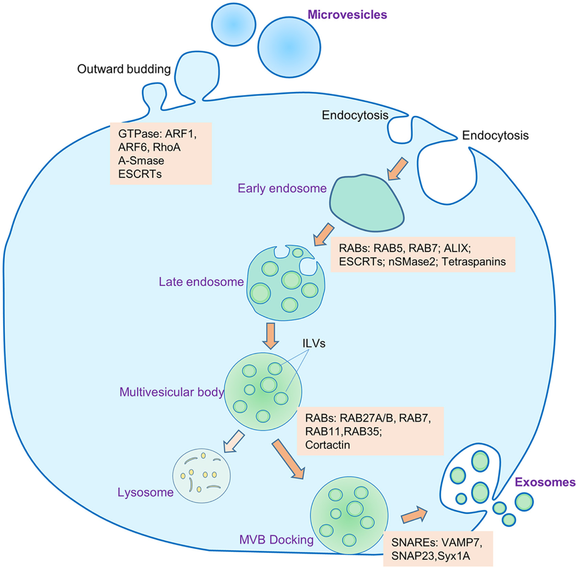
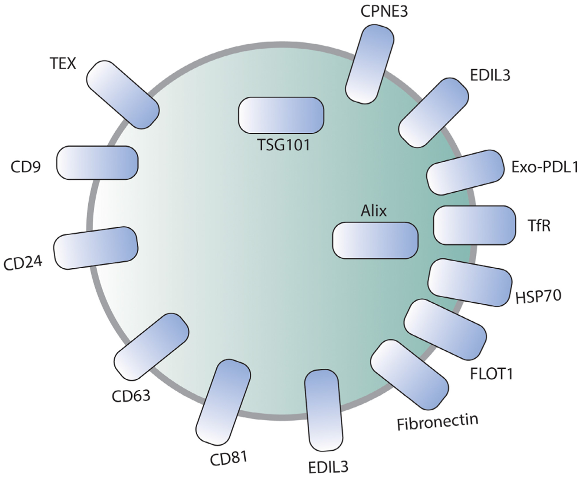
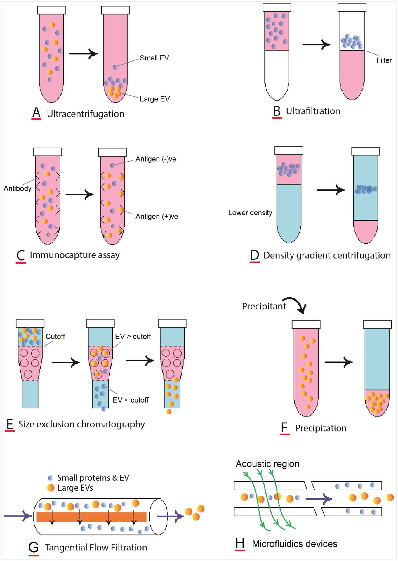
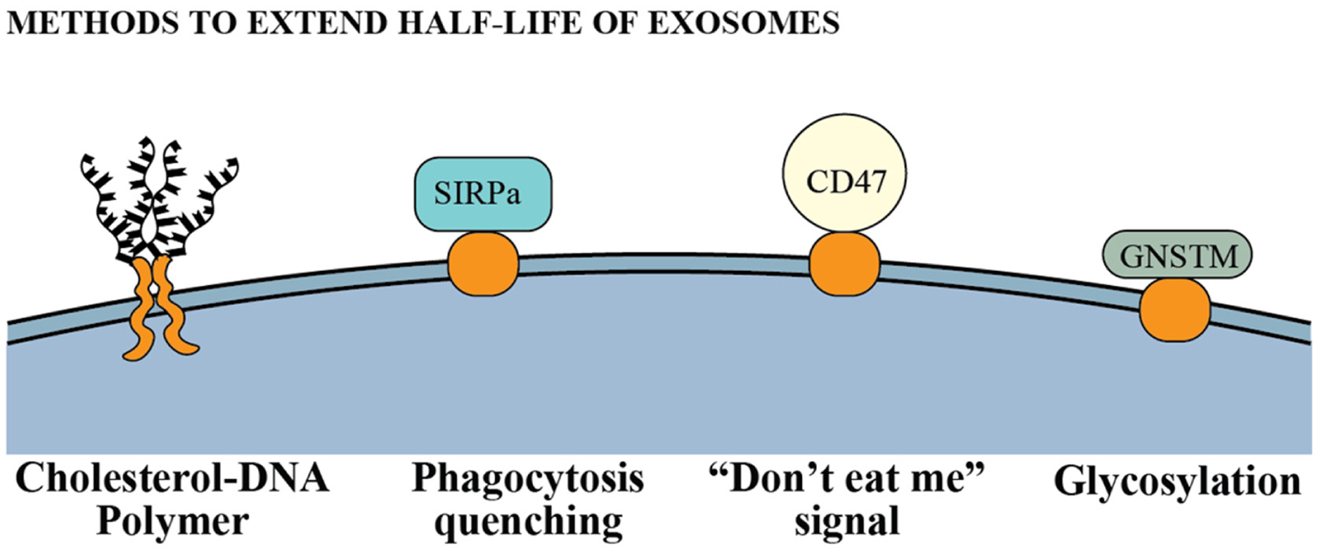
Exosomes are a type of extracellular vesicle (EV) with diameters of 30-150 nm secreted by most of the cells into the extracellular spaces and can alter the microenvironment through cell-to-cell interactions by fusion with the plasma membrane and subsequent endocytosis and release of the cargo. Because of their biocompatibility, low toxicity and immunogenicity, permeability (even through the blood-brain barrier (BBB)), stability in biological fluids, and ability to accumulate in the lesions with higher specificity, investigators have started making designer's exosomes or engineered exosomes to carry biologically active protein on the surface or inside the exosomes as well as using exosomes to carry drugs, micro RNA, and other products to the site of interest. In this review, we have discussed biogenesis, markers, and contents of various exosomes including exosomes of immune cells. We have also discussed the current methods of making engineered and designer's exosomes as well as the use of engineered exosomes targeting different immune cells in the tumors, stroke, as well as at peripheral blood. Genetic engineering and customizing exosomes create an unlimited opportunity to use in diagnosis and treatment. Very little use has been discovered, and we are far away to reach its limits.

Keywords: designer's exosomes; engineered exosomes; manipulation of biogenesis, exosomes, and immune cells; separation of exosomes; therapeutic exosomes.


Conflict of interest statement

CONFLICT OF INTEREST

None declared.

Figures

FIGURE 1
FIGURE 1
Biogenesis and secretion of exosomes and microvesicles. Microvesicles are generated by outward budding of the plasma membrane with the help of several GTPases. The process of biogenesis and release of exosomes into the extracellular space encompasses several distinct steps: (1) invagination of plasma membrane and formation of early endosomes, (2) inward protrusion of early endosomal membrane to generate late endosomes, (3) formation of multivesicular bodies (MVBs) that contain intraluminal vesicles (ILVs), (4) docking of the MVBs to the cellular plasma membrane, (5) exocytosis of the exosomes into the extracellular milieu. Some of the MVBs may go into lysosomal degradation. Several molecules are involved in the biogenesis and release of microvesicles and exosomes. ESCRT, endosome sorting complex required for transport; RAB, RAS-related protein; ALIX, ALG-2 interacting protein X; nSMase2, neutral sphingomyelinase 2; SNARE, soluble NSF attachment protein receptor; VAMP7, vesicle-associated membrane protein 7; SNAP23, synaptosomal-associated protein 23; Syx1A, syntaxin 1A; ARF, ADP, ribosylation factor; RohA, Ras homolog family member A; A-SMase, acid sphingomyelinase
FIGURE 2
FIGURE 2
Functional molecules in the exosomes released from different immune cells
FIGURE 3
FIGURE 3
Schematic diagram showing common exosomal markers
FIGURE 4
FIGURE 4
Schematic presentation of processes involved in different techniques of exosome isolation
FIGURE 5
FIGURE 5
Current methods to display a cargo on the surface of exosomes. PDGFR, Platelet-derived growth factor receptor; PTGFRN, Prostaglandin F2 Receptor Inhibitor; BLV, Bovine Leukemia Virus; LAMP2b, Lysosome-associated membrane protein 2; NGFR, Nerve Growth Factor Receptor; DSPE, 1,2-Distearoyl-snglycero-3-phosphorylethanolamine; DMPE, 1,2-Dimyristoyl-sn-glycero-3-phosphoethanolamine; DOPE, 1,2-dioleoyl-sn-glycero-3-phosphoethanolamine; PEG, Polyethylene glycol; EDC-NHS, (1-Ethyl-3-[3-dimethylami- nopropyl]-carbodiimide hydrochloride – N-Hydroxysuccinimide; N3, azide radical
FIGURE 6
FIGURE 6
Methods to load cargo inside exosomes. (A) Genetic engineering methods to load exosomes with protein and nucleic acid. MS2CP, MS2 coat protein; MBS, MS2 binding site; CRY2, cryptochrome 2; CIBN, truncated version of CRY-interaction basicloop- helix 1 protein; NLS, nuclear localization signal; BASP1, Brain Abundant Membrane Attached Signal Protein 1; HuR, Human Antigen R. (B) Physical methods to load proteins and nucleic acids into exosomes
FIGURE 7
FIGURE 7
Immunological use of engineered exosomes. CEA, carcinoembryonic antigen; HER2, human epidermal growth factor receptor 2; RSV, respiratory syncytial virus; IL6ST, Interleukin 6 Cytokine Family Signal Transducer, mIgG2b, mouse immunoglobulin G 2b
FIGURE 8
FIGURE 8
Engineering methods to extend half-life of exosomes in circulation. SIRPα, signal regulatory protein; GNSTM, glycosylation motif
FIGURE 9
FIGURE 9
(A) Disease-free and overall survival of patients with different cancers expressing mannose receptor (MRC1) in the tumor tissues (TCGA data). (B) Increased number of CD206+/CD11b+ cells in lung metastasis (middle panel, yellow cells) from breast cancer compared to that in the primary tumor (left panel). Quantitative analysis showed a significantly increased number of CD11b+CD206+ cells. The samples are from multiple patients and randomly selected histochemical sections (n = 6). * = P < 0.01
FIGURE 10
FIGURE 10
(A) Vector design to express M2 targeting peptide and Fc-mIgG2b on exosomes. (B) Cartoon to show the mechanisms of ADCC through engineered exosomes
FIGURE 11
FIGURE 11
Mobilization of neutrophils and M1 macrophages in the peripheral blood and in the stroke area was observed as early as 3 hrs. Whereas other cell types such as NK cells and macrophages (F4/80+, which also contain M2 type macrophages) gradually increased in the stroke areas. Following collection of peripheral blood from each stroke animal, animals were euthanized and perfused with ice cold PBS and the brain tissues from stroke area were collected and single cell suspensions were made for flow cytometry. * = significant differences
FIGURE 12
FIGURE 12
Survival probability in BRCA+ breast cancer patients expressing FPR1. (TCGA data from UACLAN)

Similar articles

Cited by

References

    1. van Niel G, D’Angelo G, Raposo G. Shedding light on the cell biology of extracellular vesicles. Nat Rev Mol Cell Biol. 2018;19:213. - PubMed
    1. Li X, Corbett AL, Taatizadeh E, et al. Challenges and opportunities in exosome research—Perspectives from biology, engineering, and cancer therapy. APL Bioeng. 2019;3(1):011503. - PMC - PubMed
    1. Valadi H, Ekstrom K, Bossios A, Sjostrand M, Lee JJ, Lotvall JO. Exosome-mediated transfer of mRNAs and microRNAs is a novel mechanism of genetic exchange between cells. Nat Cell Biol. 2007;9(6):654–659. - PubMed
    1. Lorentzen E, Dziembowski A, Lindner D, Seraphin B, Conti E. RNA channelling by the archaeal exosome. EMBO Rep. 2007;8(5):470–476. - PMC - PubMed
    1. Denzer K, Kleijmeer MJ, Heijnen HF, Stoorvogel W, Geuze HJ. Exosome: from internal vesicle of the multivesicular body to intercellular signaling device. J Cell Sci. 2000;113(Pt 19):3365–3374. - PubMed

Publication types