. 2022 Sep 28;107(10):e4037-e4047.
doi: 10.1210/clinem/dgac415.

Teprotumumab Divergently Alters Fibrocyte Gene Expression: Implications for Thyroid-associated Ophthalmopathy

Affiliations

Teprotumumab Divergently Alters Fibrocyte Gene Expression: Implications for Thyroid-associated Ophthalmopathy

Roshini Fernando et al. J Clin Endocrinol Metab. .

Abstract

Context: Teprotumumab, an IGF-I receptor (IGF-IR) inhibitor, is effective in thyroid-associated ophthalmopathy (TAO). The drug can modulate induction by TSH of IL-6 and IL-8 in CD34+ fibrocytes and their putative derivatives, CD34+ orbital fibroblasts (CD34+ OF). Fibrocytes express multiple thyroid autoantigens and cytokines implicated in TAO, which are downregulated by Slit2. Inflammation and disordered hyaluronan (HA) accumulation occur in TAO. Whether teprotumumab alters these processes directly in fibrocytes/CD34+ OF remains uncertain.

Objective: Determine teprotumumab effects on expression/synthesis of several TAO-relevant molecules in fibrocytes and GD-OF.

Design/setting/participants: Patients with TAO and healthy donors were recruited from an academic endocrine and oculoplastic practice.

Main outcome measures: Real-time PCR, specific immunoassays.

Results: Teprotumumab attenuates basal and TSH-inducible autoimmune regulator protein, thyroglobulin, sodium iodide symporter, thyroperoxidase, IL-10, and B-cell activating factor levels in fibrocytes. It downregulates IL-23p19 expression/induction while enhancing IL-12p35, intracellular and secreted IL-1 receptor antagonists, and Slit2. These effects are mirrored by linsitinib. HA production is marginally enhanced by teprotumumab, the consequence of enhanced HAS2 expression.

Conclusion: Teprotumumab affects specific gene expression in fibrocytes and GD-OF in a target-specific, nonmonolithic manner, whereas IGF-IR control of these cells appears complex. The current results suggest that the drug may act on cytokine expression and HA production systemically and locally, within the TAO orbit. These findings extend our insights into the mechanisms through which IGF-IR inhibition might elicit clinical responses in TAO, including a potential role of Slit2 in attenuating inflammation and tissue remodeling.

Keywords: Graves’ disease; cytokine; fibroblasts; hyaluronan; ophthalmopathy; orbit.


Figures

Figure 1.
Figure 1.
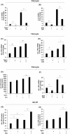
Effect of teprotumumab on bTSH-induced thyroid-specific genes Tg, TPO, NIS, and AIRE in fibrocytes. Confluent fibrocyte cultures were treated with or without teprotumumab (50 µg/mL) or human isotype IgG for 7-9 days, followed by bTSH (5 mIU/mL) for 6 hours. Monolayers were washed with PBS and RNA was extracted and reverse transcribed. Harvested cDNA was subjected to quantitative real-time PCR for (A) AIRE, (B) Tg, (C) TPO, and (D) NIS mRNA levels. Values were normalized to their respective GAPDH levels and are expressed as the mean ± SD of 3 independent determinations. Experiments were repeated 3 times. *P < 0.05; **P < 0.01.
Figure 2.
Figure 2.
Divergent effect of teprotumumab on bTSH induced IL-23p19 and IL-12p35 expression on fibrocyte and GD-OF. Rinsed fibrocyte and GD-OF monolayers were incubated with or without (A,B) teprotumumab (50 µg/mL) or (C,D) linsitinib (1 µM) and human isotype IgG for 7-9 days, and bTSH (5 mIU/mL) was added for 6 hours. Harvested RNA was reverse transcribed and isolated cDNA was subjected to quantitative real-time PCR for IL-23p19 and IL-12p35 mRNA levels. Values were normalized to their respective GAPDH levels and are expressed as the mean ± SD of 3 independent determinations. Experiments were repeated 3 times. **P < 0.01; ***P < 0.001.
Figure 3.
Figure 3.
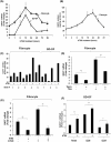
Teprotumumab attenuates bTSH induced IL-10 while enhancing icIL-1RA and sIL-1RA expression in fibrocytes and GD-OF. Fibrocyte cultures were treated with teprotumumab (50 µg/mL), rhSlit2 (50 ng/mL) or human isotype IgG for 7-9 days, with bTSH (5 mIU/mL) for the final 6 hours. (A-F) RNA was extracted and reverse transcribed. Isolated cDNA was subjected to quantitative real-time PCR for (A,B) IL-10, (C,D) icIL-1RA, (F) siIL-1RA. (E,G,H) Media and cellular proteins were collected and subjected to specific ELISAs for icIL-1RA and sIL-1RA. PCR values were normalized to their respective GAPDH levels. icIL-1RA and sIL-1RA protein levels were normalized to cellular protein levels. Data are expressed as the mean ± SD of 3 independent determinations. Experiments were repeated 3 times. **P < 0.01; ***P < 0.001.
Figure 4.
Figure 4.
Effect of bTSH, teprotumumab, and Slit2 on BAFF expression in fibrocytes and GD-OF. (A,B) Cultures were treated for the graded intervals indicated along the abscissas with bTSH (5 mIU/mL). (A) Cellular RNA was harvested and subjected to reverse transcription by RT-PCR. Values were normalized to their respective GAPDH levels (B) Collected medium was assayed for BAFF levels with an ELISA, described in Methods. Values were normalized to respective monolayer protein content. Data are expressed as mean ± SD of triplicate determinations; *P < 0.05; **P < 0.01; ***P < 0.001. (C-F), bTSH for (12 hours), or teprotumumab (50 µg/mL) or rhSlit2 (50 ng/mL) was added for 7 days and analyzed for BAFF mRNA levels as in panel A. (C) Multiple culture strains were left untreated (-) or treated with bTSH for 12 hours. (D,E) Cultures of fibrocytes were treated with nothing, bTSH (12 hours), teprotumumab, or Slit2 or the combinations indicated. (F) Parental GD-OF strains were sorted into pure CD34+ and CD34 subsets using a FACSAria III instrument. Subsets were then cultured for 48 hour, the final 12 hours in the absence or presence of bTSH. Data were expressed as mean ± SD of triplicate independent determinations from a single experiment representative of 3 conducted. *P < 0.05; **P < 0.01.
Figure 5.
Figure 5.
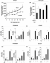
Effect of teprotumumab on bTSH-induced HA production in (A) GD-OF and (B) fibrocytes. GD-OF were treated with or without teprotumumab (50 µg/mL) or human isotype IgG (5 µg/mL) for 7-9 days and stimulated with bTSH (5 mIU/mL) for graded intervals indicated along the abscissa for 0 to 96 hours. Media were collected and subjected to HA ELISA. Values were normalized to respective cell layer protein levels and expressed as the mean ± SD of 3 independent determinations. Experiments were repeated 3 times. (C-F) Divergent pattern of HAS isoenzyme induction by bTSH in fibrocytes versus GD-OF and effects of (C,D) teprotumumab and (E,F) linsitinib. Confluent monolayers of GD-OF and fibrocytes remained untreated or received teprotumumab (50 µg/mL) or linsitinib (1 µM) and human Isotype IgG for 7-9 days. Some cultures were treated with bTSH (5 mIU/mL) for 6 hours. Cellular RNA was harvested, mRNA was reverse transcribed, and cDNA subjected to quantitative real-time PCR for HAS1 and HAS2. Values were normalized to their respective GAPDH levels and are expressed as the mean ± SD of 3 independent determinations. Experiments were repeated 3 times. *P < 0.05; **P < 0.01; ***P < 0.001.
Figure 6.
Figure 6.
Teprotumumab induces Slit2 and ROBO1 mRNA and Slit2 protein in GD-OF. Cultures were treated with either isotype control IgG or teprotumumab (50 µg/mL) for 3 days. (A) Culture media were collected and subjected to Slit2-specific ELISA. Levels of Slit2 were normalized to respective cell layer protein content. (B) Cellular RNA was harvested from rinsed cell layers and mRNA reverse transcribed, and cDNA subjected to quantitative real-time PCR for Slit2 and ROBO1. Values were normalized to their respective GAPDH levels. **P < 0.01, ***P < 0.001.

Similar articles

Cited by

References

    1. Kazim M, Goldberg RA, Smith TJ. Insights into the pathogenesis of thyroid-associated orbitopathy: evolving rationale for therapy. Arch Ophthalmol. 2002;120(3):380-386. - PubMed
    1. Smith TJ, Hegedus L. Graves’ disease. N Engl J Med. 2016;375(6):1552-1565. - PubMed
    1. Feliciello A, Porcellini A, Ciullo I, Bonavolonta G, Avvedimento EV, Fenzi G. Expression of thyrotropin-receptor mRNA in healthy and Graves’ disease retro-orbital tissue. Lancet. 1993;342(8867):337-338. - PubMed
    1. Heufelder AE, Dutton CM, Sarkar G, Donovan KA, Bahn RS. Detection of TSH receptor RNA in cultured fibroblasts from patients with Graves’ ophthalmopathy and pretibial dermopathy. Thyroid. 1993;3(4):297-300. - PubMed
    1. Smith TJ, Hoa N. Immunoglobulins from patients with Graves’ disease induce hyaluronan synthesis in their orbital fibroblasts through the self-antigen, insulin-like growth factor-I receptor. J Clin Endocrinol Metab. 2004;89(10):5076-5080. - PubMed

Publication types

MeSH terms