. 2022 Aug 30;434(16):167706.
doi: 10.1016/j.jmb.2022.167706. Epub 2022 Jul 6.

Structural Basis of Main Proteases of Coronavirus Bound to Drug Candidate PF-07304814

Affiliations

Structural Basis of Main Proteases of Coronavirus Bound to Drug Candidate PF-07304814

Jian Li et al. J Mol Biol. .

Abstract

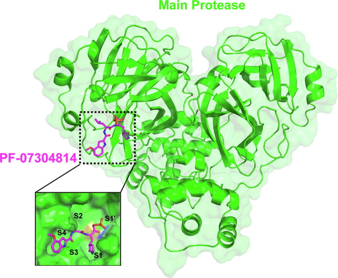
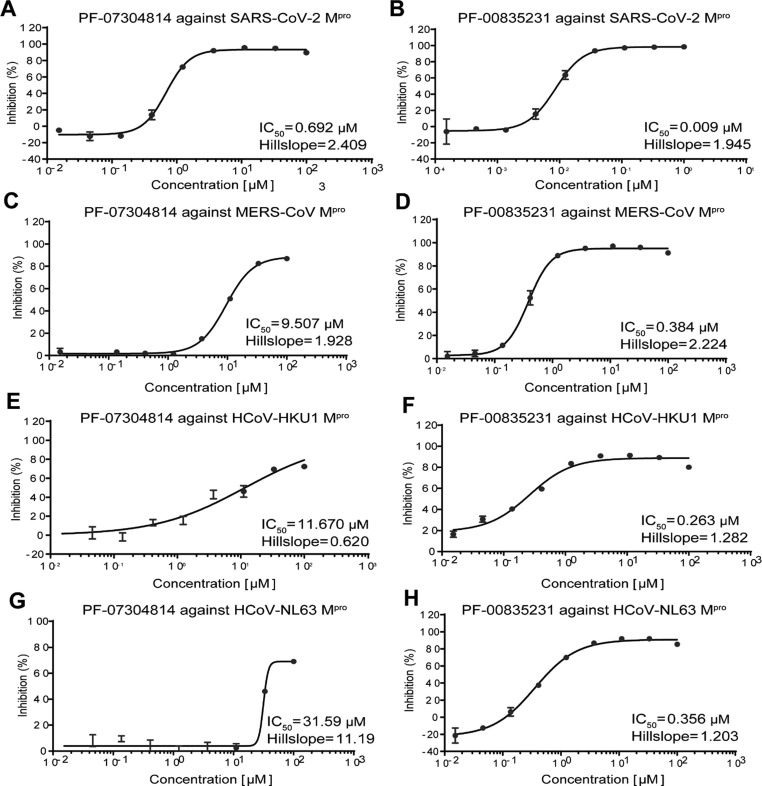
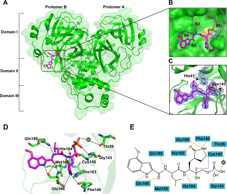
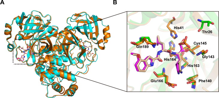
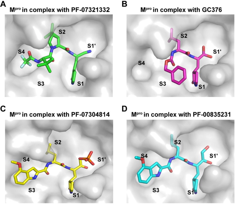
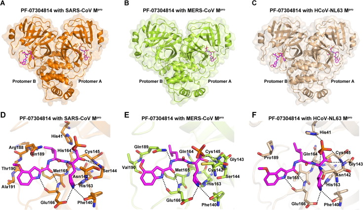
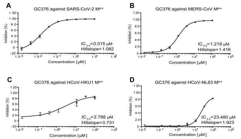
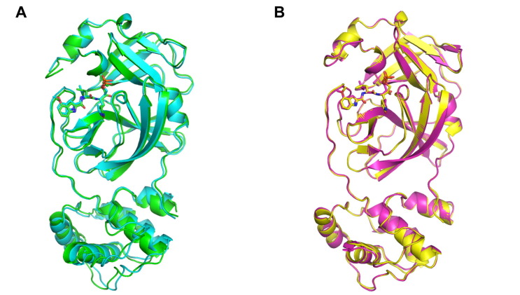
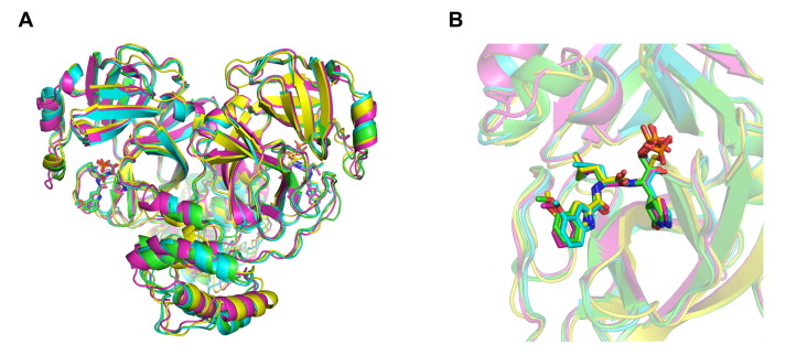
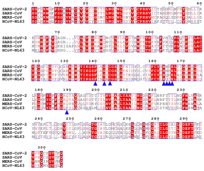
New variants of the severe acute respiratory syndrome Coronavirus 2 (SARS-CoV-2) emerged and spread rapidly all over the world, which strongly supports the need for pharmacological options to complement vaccine strategies. Main protease (Mpro or 3CLpro) is a critical enzyme in the life cycle of SARS-CoV-2 and appears to be highly conserved among different genera of coronaviruses, making it an ideal target for the development of drugs with broad-spectrum property. PF-07304814 developed by Pfizer is an intravenously administered inhibitor targeting SARS-CoV-2 Mpro. Here we showed that PF-07304814 displays broad-spectrum inhibitory activity against Mpros from multiple coronaviruses. Crystal structures of Mpros of SARS-CoV-2, SARS-CoV, MERS-CoV, and HCoV-NL63 bound to the inhibitor PF-07304814 revealed a conserved ligand-binding site, providing new insights into the mechanism of inhibition of viral replication. A detailed analysis of these crystal structures complemented by comprehensive comparison defined the key structural determinants essential for inhibition and illustrated the binding mode of action of Mpros from different coronaviruses. In view of the importance of Mpro for the medications of SARS-CoV-2 infection, insights derived from the present study should accelerate the design of pan-coronaviral main protease inhibitors that are safer and more effective.

Keywords: PF-07304814; coronavirus; inhibitor; main protease; structural basis.


Conflict of interest statement

Conflict of interest The authors declare no conflict of interest.

Figures

None
Graphical abstract
Figure 1
Figure 1
Enzymatic inhibition of PF-07304814 and PF-00835231 against main proteasesof different coronaviruses. Inhibition of PF-07304814 and PF-00835231 against main protease of SARS-CoV-2 (A, B), MERS-CoV (C, D), HCoV-HKU1 (E, F) and HCoV-NL63 (G, H). Main proteases were preincubated in the reaction buffer with various concentrations of PF-07304814 or PF-00835231 at room temperature for 30 min before reacting with the FRET substrate The IC50 values were calculated using the GraphPad Prism software.
Figure 2
Figure 2
Crystal structure of SARS-CoV-2 Mpro in complex with PF-07304814. (A) Overall structure of SARS-CoV-2 Mpro in complex with PF-07304814. Three domains and two protomers of Mpro are labeled. The substrate-binding pocket is situated within the black dotted box. PF-07304814 is shown as sticks with the carbon atoms in magentas, oxygen atoms in red, nitrogen atoms in blue, and phosphorus atoms in orange. (B) An enlarged view of the substrate-binding pocket. PF-07304814 forms a covalent bond with Cys145. The substrate-binding subsites (S1′, S1, S2, S3 and S4) are labeled. (C) A C-S covalent bond forms between Cys145 and the hydroxymethyl ketone of PF-07304814. The 2Fo–Fc density map contoured at 1.0σ is shown as blue mesh. (D) The detailed interaction in the complex structure is shown with the residues involved in inhibitor binding (within 3.5 Å) displayed as sticks. W represents the water molecule. Hydrogen-bonding interactions are shown as black dashed lines. (E) Schematic interaction between PF-07304814 and Mpro. Hydrogen-bonding interactions are shown as orange dashed lines.
Figure 3
Figure 3
Comparison of the binding modes of PF-07304814 and PF-00835231. (A) Comparison of overall structures between SARS-CoV-2 Mpro-PF-07304814 and SARS-CoV-2 Mpro-PF-00835231 (PDB ID 6XHM). Structures of SARS-CoV-2 Mpro bound to PF-07304814 and PF-00835231 are shown in cyan and orange, respectively. PF-07304814 and PF-00835231 are shown in magentas and wheat sticks, respectively. (B) A zoomed in view of substrate binding pocket of SARS-CoV-2 Mpro. The key residues of SARS-CoV-2 Mpro involved in hydrogen-bonding interaction with PF-07304814 and PF-00835231 are shown as green and orange sticks, respectively.
Figure 4
Figure 4
Comparison of the binding modes of four inhibitors targeting SARS-CoV-2 Mpro. (A to D) The binding pockets of PF-07321332 (A) (PDB ID 7VLQ), GC376 (B) (PDB ID 7D1M), PF-07304814 (C) (PDB ID 7VVP), and PF-00835231 (D) (PDB ID 6XHM) bound to SARS-CoV-2 Mpro are shown. Mpros are shown as the gray surface, and the inhibitors are shown as sticks.
Figure 5
Figure 5
Crystal structures of SARS-CoV, MERS-CoV and HCoV-NL63 Mpros in complex with PF-07304814. (A–C) Overall structures of SARS-CoV Mpro (A), MERS-CoV Mpro (B) and HCoV-NL63 Mpro (C) in complex with PF-07304814. Two protomers of Mpro are labeled. PF-07304814 is shown as sticks with the carbon atoms in magentas, oxygen atoms in red, nitrogen atoms in blue, and phosphorus atoms in orange. (D-F) The detailed interactions in the SARS-CoV Mpro-PF-07304814 complex (D), MERS-CoV Mpro-PF-07304814 complex (E), and HCoV-NL63 Mpro-PF-07304814 complex (F) are shown with the residues involved in inhibitor binding (within 3.5 Å) displayed as sticks. Hydrogen-bonding interactions are shown as black dashed lines.
Supplementary Figure S1
Enzymatic inhibition of GC376 against main protease of different coronaviruses. Inhibition of GC376 against main protease of SARS-CoV-2 (A), MERS-CoV (B), HCoV-HKU1 (C), and HCoV-NL63 (D). Main proteases were preincubated in the reaction buffer with various concentrations of GC376 at room temperature for 30 min before reacting with the FRET substrate The IC50 values was calculated using the GraphPad Prism software.
Supplementary Figure S2
Structural comparison of protomer A and B of SARS-CoV and HCoV-NL63 Mpros bound to PF-07304814. (A) Structural comparison of protomer A and protomer B of SARS-CoV Mpro bound to PF-07304814. Protomer A (cyan) and B (green) of SARS-CoV Mpro are shown as cartoon, while ligand shown as sticks. (B) Structural comparison of protomer A and protomer B of HCoV-NL63 Mpro bound to PF-07304814. Protomer A (yellow) and B (magentas) of HCoV-NL63 Mpro are shown as cartoon, while ligand shown as sticks.
Supplementary Figure S3
Structural comparison of PF-07304814 in complex with Mpros of SARS-CoV, MERS-CoV, and HCoV-NL63. (A) Structural alignment of CoV Mpros bound to PF-07304814 with SARS-CoV-2 Mpro-inhibitor complex in green, SARS-CoV Mpro-inhibitor complex in cyan, MERS-CoV Mpro-inhibitor complex in yellow, and HCoV-NL63 Mpro-inhibitor complex in magentas. (B) An enlarged view of the substrate-binding pocket.
Supplementary Figure S4
Sequence alignment of Mpros from SARS-CoV-2, SARS-CoV, MERS-CoV and HCoV-NL63. Amino acid sequence of Mpros from SARS-CoV-2, SARS-CoV, MERS-CoV and HCoV-NL63 are retrieved from GenBank. ClustalX (1.83) software is used for sequence alignment and ESPript 3.0 was used to generate a graphical representation. The key residues involve in interacting with inhibitors are labeled with blue triangle.

References

    1. Zhou P., Yang X.L., Wang X.G., Hu B., Zhang L., Zhang W., Si H.R., Zhu Y., et al. A pneumonia outbreak associated with a new coronavirus of probable bat origin. Nature. 2020;579:270–273. - PMC - PubMed
    1. Zhu N., Zhang D., Wang W., Li X., Yang B., Song J., Zhao X., Huang B., et al. A Novel Coronavirus from Patients with Pneumonia in China, 2019. N. Engl. J. Med. 2020;382:727–733. - PMC - PubMed
    1. Ye Z.W., Yuan S., Yuen K.S., Fung S.Y., Chan C.P., Jin D.Y. Zoonotic origins of human coronaviruses. Int. J. Biol. Sci. 2020;16:1686–1697. - PMC - PubMed
    1. Su S., Wong G., Shi W., Liu J., Lai A.C.K., Zhou J., Liu W., Bi Y., et al. Epidemiology, Genetic Recombination, and Pathogenesis of Coronaviruses. Trends Microbiol. 2016;24:490–502. - PMC - PubMed
    1. Chan J.F., Kok K.H., Zhu Z., Chu H., To K.K., Yuan S., Yuen K.Y. Genomic characterization of the 2019 novel human-pathogenic coronavirus isolated from a patient with atypical pneumonia after visiting Wuhan. Emerg. Microbes Infect. 2020;28:221–236. - PMC - PubMed

Publication types

LinkOut - more resources