

Review
doi: 10.1016/j.cell.2022.06.016.
Epub 2022 Jul 8.
Tools for mammalian glycoscience research
Affiliations
- PMID: 35809571
- PMCID: PMC9339253
- DOI: 10.1016/j.cell.2022.06.016
Item in Clipboard
Review
Tools for mammalian glycoscience research
Cell.
.
Abstract
Cellular carbohydrates or glycans are critical mediators of biological function. Their remarkably diverse structures and varied activities present exciting opportunities for understanding many areas of biology. In this primer, we discuss key methods and recent breakthrough technologies for identifying, monitoring, and manipulating glycans in mammalian systems.
Keywords: carbohydrates; glycans; glycobiology; mammalian biology.
Copyright © 2022 Elsevier Inc. All rights reserved.
Conflict of interest statement
Declaration of interests The authors declare no competing interests.
Figures
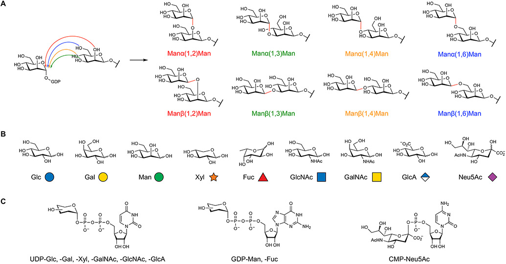
(A) The native assembly of two monosaccharides leads to eight potential disaccharides, depending on the regioselectivity (e.g., 1,2 versus 1,3) and stereoselectivity (α versus β) of the newly formed glycosidic bond. In contrast, only a single structure is produced from the native assembly of dinucleotides or dipeptides. (B) Mammalian glycans are composed of nine monosaccharides, which are pictorially represented by specific symbols from the Symbol Nomenclature for Glycans. (C) Glycosyltransferases utilize activated nucleotide sugar donors to transfer monosaccharide units onto growing glycan chains.
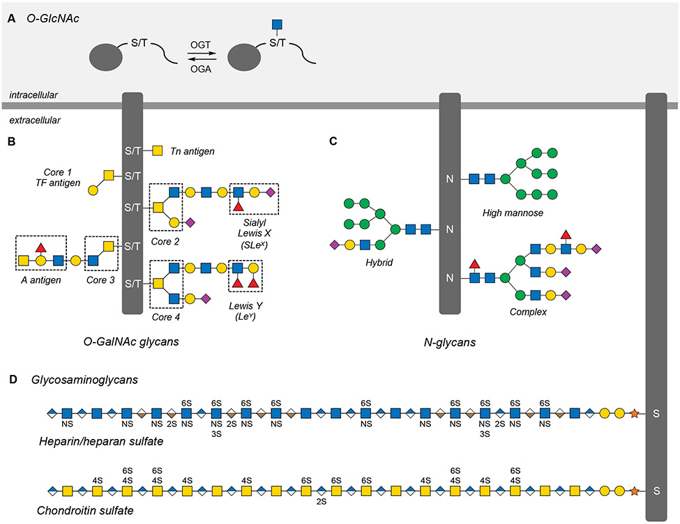
(A) O-GlcNAcylation is the dynamic and reversible addition of N-acetylglucosamine to Ser and Thr residues on thousands of intracellular proteins. (B) O-GalNAc or mucin-like glycans are a broad class of O-linked extracellular glycans categorized by one of eight core structures that can be elaborated with a number of glycan antigens. (C) N-glycans are branched glycans attached to Asn residues of extracellular proteins and are categorized by the number and composition of their antennae branching from a conserved core structure. (D) Glycosaminoglycans (GAGs) are linear extracellular polysaccharides that can be sulfated at different hydroxyl and amine positions along the length of the glycan chain.
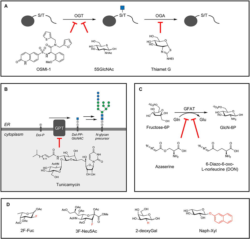
(A) Protein O-GlcNAcylation can be blocked through the OGT inhibitors OSMI-1 and 5SGlcNAc, and the removal of O-GlcNAc can be targeted through the OGA inhibitor Thiamet G. (B) N-glycosylation can be broadly inhibited using tunicamycin, which inhibits the attachment of GlcNAc-1-phosphate to dolichol phosphate (Dol-P) by GlcNAc-1-phosphate transferase (GPT). (C) Glycans containing GlcNAc and GalNAc can be targeted using the glutamine mimics azaserine and DON, which target GFAT activity in hexosamine biosynthesis. (D) “Look-alike” mimics of monosaccharides can inhibit specific modifications such as fucosylation (2F-Fuc and 2-deoxyGal) and sialylation (3F-Neu5Ac) or act as decoys for GTs in GAG biosynthesis (Naph-Xyl).
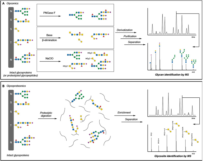
(A) Glycomics, or the analysis of glycan composition, is accomplished through the enzymatic or chemical release of glycans, followed by chemical derivatization, purification, separation, and mass spectrometric characterization of glycan structures. (B) Glycoproteomics, or the analysis of protein glycosylation, is accomplished through the digestion of glycoproteins and enrichment of glycopeptides, followed by separation and mass spectrometric identification of glycosylated peptides.
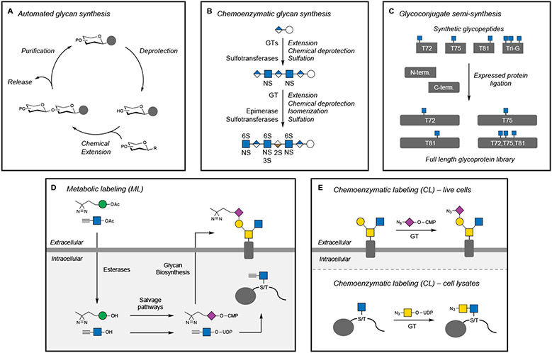
(A) Automated methods for the chemical synthesis of oligosaccharides use a similar approach as solid-phase peptide or oligonucleotide synthesis. The glycan is elongated through a series of deprotection and coupling steps, followed by release from the resin. (B) The chemoenzymatic synthesis of an Arixtra biosimilar oligosaccharide employs multiple enzymatic and chemical steps to assemble and functionalize an HS heptasaccharide. (C) Semi-synthesis of O-GlcNAcylated α-synuclein utilizes synthetic glycopeptide fragments for expressed protein ligation to generate a library of specific protein glycoforms. (D) Metabolic labeling (ML) utilizes peracetylated (indicated by OAc) nonnatural monosaccharides that cross cell membranes, are deprotected (indicated by OH) by endogenous esterases, converted into nucleotide sugar donors, and then incorporated by GTs into glyconjugates. Nonnatural glycans with diazirine or alkyne functionalities are shown as representative examples. (E) Chemoenzymatic labeling (CL) employs exogenous GTs and nonnatural nucleotide sugar donors to modify specific glycan structures recognized by the GT. Nonnatural glycans with azide functionalities are shown.
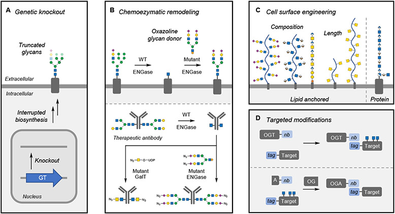
(A) Genetic knockout of individual glycosyltransferases (GTs) and other glycan biosynthetic enzymes globally affects cellular glycan populations, leading to truncated glycan structures. (B) Chemoenzymatic remodeling using endoglycosidases (ENGase) simplifies N-glycan heterogeneity on the cell surface or on proteins. Installation of specific structures is accomplished using mutant ENGases or GTs that can install specific structures functionalized for further biorthogonal reactions. (C) Cell-surface engineering has been accomplished with both synthetic and naturally occurring glycopolymers anchored to the cell surface by lipids or proteins. Techniques can modulate the glycocalyx by changing its composition or thickness. (D) Targeted O-GlcNAc modification utilizes nanobody (nb)-fused OGT or OGA, which directs the enzymes to tagged target proteins. In the case of OGA, this approach is accomplished through a split OGA construct.
References
-
- Amon R, Grant OC, Leviatan Ben-Arye S, Makeneni S, Nivedha AK, Marshanski T, Norn C, Yu H, Glushka JN, Fleishman SJ, et al. (2018). A combined computational-experimental approach to define the structural origin of antibody recognition of sialyl-Tn, a tumor-associated carbohydrate antigen. Sci Rep 8,10786. 10.1038/s41598-018-29209-9. - DOI - PMC - PubMed
Publication types
MeSH terms
Substances
Grants and funding
LinkOut - more resources
Full Text Sources
