. 2022 Jul 9;9(1):66.
doi: 10.1186/s40634-022-00501-y.

Developing an experimental model of early knee osteoarthritis after medial meniscus posterior root release: an in vivo study

Affiliations

Developing an experimental model of early knee osteoarthritis after medial meniscus posterior root release: an in vivo study

Lika Dzidzishvili et al. J Exp Orthop. .

Abstract

Purpose: To develop a predictable and reproducible model of knee osteoarthritis after medial meniscus posterior root release.

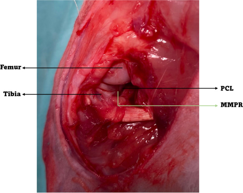
Methods: Posteromedial meniscal root tears were created in 12 White New Zealand rabbit knees. The contralateral limbs were used as healthy controls. The animals were euthanized at 16 weeks postoperatively; tissue samples of femoral and tibial articular cartilage were collected and processed for macro and microscopic analyses to detect signs of early degeneration. Clinical evaluation of the weight-bearing status on the affected knee was conducted at 0-, 4-, 8-, and 16-weeks postoperatively.

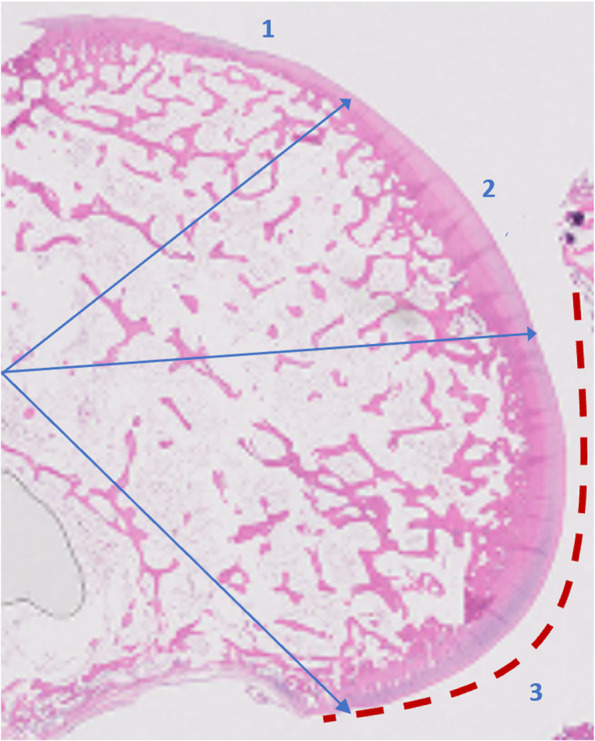
Results: Early and severe osteoarthritic changes were the hallmark and the main findings after 16-weeks post-surgery. Macroscopically, extensive osteoarthritic changes were observed across the femoral condyle and tibial plateau. Microscopic finding included ulcerations, fissures, fibrillations, pitting, and loss of the superficial layer. Cellularity was diminished, the normal pattern of distribution in columns was lost, and subchondral bone exposure was also evident.

Conclusions: This study describes a novel model of knee osteoarthritis that may guide the development of tailored interventions to delay or prevent knee osteoarthritis. This knowledge could shift the current treatment paradigm toward more conservative and knee salvageable treatment options and increase surgeons' awareness of this injury pattern. Such considerations may have a positive impact on clinical decision-making and subsequent patient-reported clinical outcomes.

Design: Controlled laboratory study.

Level of evidence: II.

Keywords: Animal model; Early osteoarthritis; Knee osteoarthritis; Meniscal root tear; Rabbit model.


Conflict of interest statement

The authors declared no potential conflicts of interest with respect to the research, authorship, and/or publication of this article.

Figures

Fig. 1
Fig. 1
Meniscectomy was performed by transecting the medial meniscus posterior root with a radial cut (PCL, posterior cruciate ligament; MMPR, medial meniscus posterior root)
Fig. 2
Fig. 2
Representative image of meniscal root remnant after 16-weeks of partial meniscectomy
Fig. 3
Fig. 3
Sagittal section of medial femoral condyles was divided in three zone of interest (1. anterior, 2. central, and 3. posterior). The red dotted line shows the weight-bearing area of the medial femoral condyle
Fig. 4
Fig. 4
Gross pathological macrographs after 16-weeks from surgery where the load bearing area of femoral condyles appears eburnated, irregularities and pitting can be detected (red arrow, 4A). Note full-thickness ulcer in the weight bearing area of the medial femoral condyle with irregularities and pitting (red arrow, 4B). The rim of the medial tibial plateau shows a whitish osteophytes and erosions (4C). Images were acquired using a 10_objective lens under bright field. Images were then digitally overlaid
Fig. 5
Fig. 5
Hematoxylin eosin (5A, 5B) and alcian blue staining (5C) of articular cartilage samples. The cartilage shows a normal appearance, the surface is smooth, chondrocytes are present throughout, and the matrix staining is homogeneous in the control knee (5A). The superficial and intermediate layers of cartilage are lost, and matrix vertical fissures reach the deep radial zone in this sample corresponding to an ulcer at the weight bearing area from an OA knee in animals subjected to meniscal root injury and partial meniscectomy (5 B, C). Cellularity is diminished and the normal pattern of distribution in columns is lost. Cartilage matrix is loss with delamination and excavation of superficial and mid layer (5B, C). Marked structural abnormalities and hypocellularity are also evident. Matrix staining intensity is severely reduced (× 20)
Fig. 6
Fig. 6
Alcian blue staining of weight bearing articular cartilage sample showing reduction of matrix staining intensity and breached tidemark by blood vessels and with minor splits (red arrows) compromising the integrity of the junction between the calcified and the noncalcified cartilage
Fig. 7
Fig. 7
Comparative radiographic images just before (9A) and after 16-weeks post-surgery (9B) showing medial joint space narrowing

References

    1. Cinque ME, Chahla J, Moatshe G, Faucett SC, Krych AJ, LaPrade RF. Meniscal root tears: a silent epidemic. Br J Sports Med. 2018;52(13):872–876. doi: 10.1136/bjsports-2017-098942. - DOI - PubMed
    1. Allaire R, Muriuki M, Gilbertson L, Harner CD. Biomechanical consequences of a tear of the posterior root of the medial meniscus. J Bone Joint Surg Am. 2008;90(9):1922–1931. doi: 10.2106/JBJS.G.00748. - DOI - PubMed
    1. Baratz ME, Fu FH, Mengato R. Meniscal tears: the effect of meniscectomy and of repair on intraarticular contact areas and stress in the human knee. A preliminary report. Am J Sports Med. 1986;14(4):270–275. doi: 10.1177/036354658601400405. - DOI - PubMed
    1. Luther JK, Cook CR, Cook JL. Meniscal release in cruciate ligament intact stifles causes lameness and medial compartment cartilage pathology in dogs 12 weeks postoperatively. Vet Surg. 2009;38(4):520–529. doi: 10.1111/j.1532-950X.2009.00520.x. - DOI - PubMed
    1. Duan X, Rai MF, Holguin N, Silva MJ, Patra D, Liao W, Sandell LJ. Early changes in the knee of healer and non-healer mice following non-invasive mechanical injury. J Orthop Res. 2017;35(3):524–536. doi: 10.1002/jor.23413. - DOI - PMC - PubMed