. 2022 Jul 10;29(1):50.
doi: 10.1186/s12929-022-00834-x.

A dorsal CA2 to ventral CA1 circuit contributes to oxytocinergic modulation of long-term social recognition memory

Affiliations

A dorsal CA2 to ventral CA1 circuit contributes to oxytocinergic modulation of long-term social recognition memory

Tsung-Chih Tsai et al. J Biomed Sci. .

Abstract

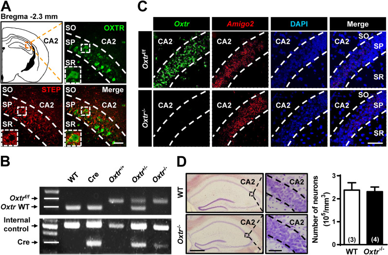
Background: Social recognition memory (SRM) is the ability to distinguish familiar from novel conspecifics and is crucial for survival and reproductive success across social species. We previously reported that oxytocin (OXT) receptor (OXTR) signaling in the CA2/CA3a of dorsal hippocampus is essential to promote the persistence of long-term SRM, yet how the endogenous OXT system influences CA2 outputs to regulate long-term SRM formation remains unclear.

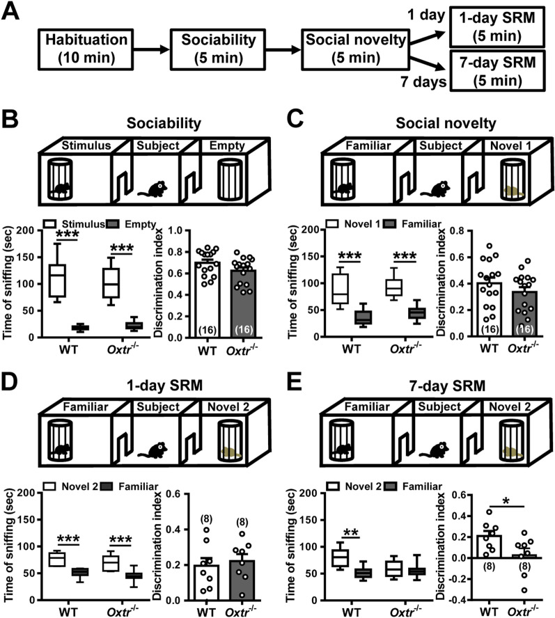
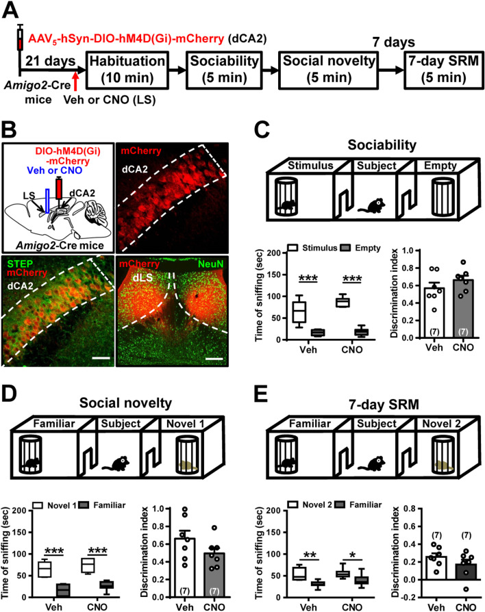
Methods: To achieve a selective deletion of CA2 OXTRs, we crossed Amigo2-Cre mice with Oxtr-floxed mice to generate CA2-specific Oxtr conditional knockout (Oxtr-/-) mice. A three-chamber paradigm test was used for studying SRM in mice. Chemogenetic and optogenetic targeting strategies were employed to manipulate neuronal activity.

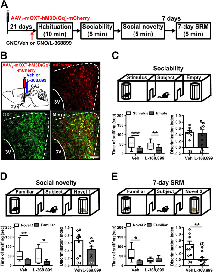
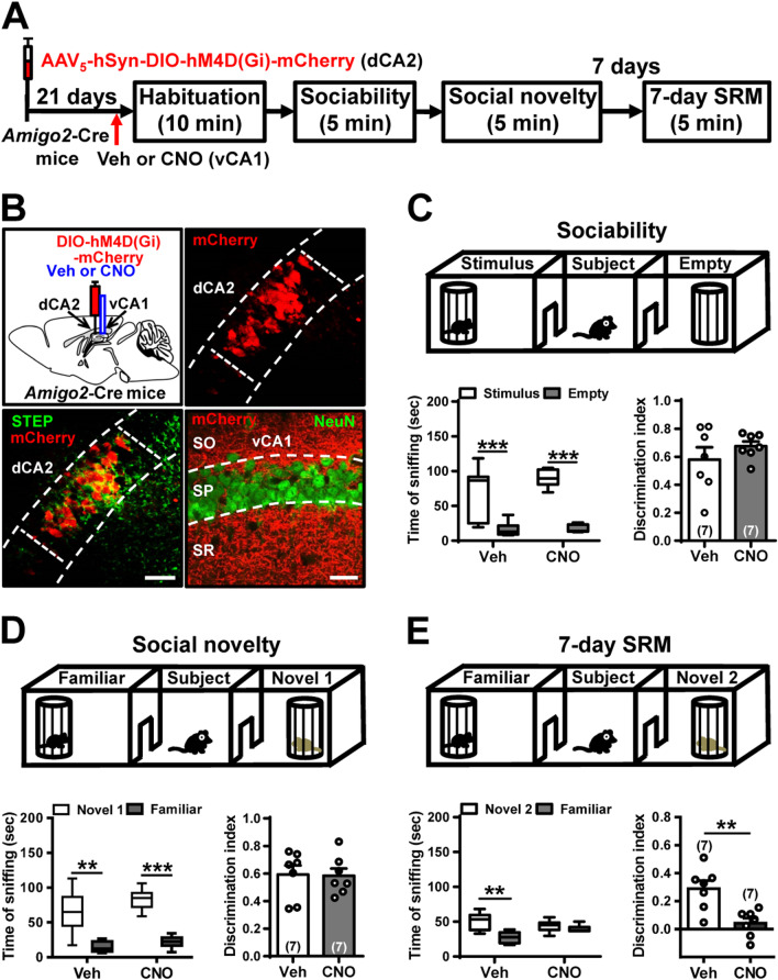
Results: We show that selective ablation of Oxtr in the CA2 suffices to impair the persistence of long-term SRM but has no effect on sociability and social novelty preference in the three-chamber paradigm test. We find that cell-type specific activation of OXT neurons within the hypothalamic paraventricular nucleus enhances long-term SRM and this enhancement is blocked by local application of OXTR antagonist L-368,899 into dorsal hippocampal CA2 (dCA2) region. In addition, chemogenetic neuronal silencing in dCA2 demonstrated that neuronal activity is essential for forming long-term SRM. Moreover, chemogenetic terminal-specific inactivation reveals a crucial role for dCA2 outputs to ventral CA1 (vCA1), but not dorsal lateral septum, in long-term SRM. Finally, targeted activation of the dCA2-to-vCA1 circuit effectively ameliorates long-term SRM deficit observed in Oxtr-/- mice.

Conclusions: These findings highlight the importance of hippocampal CA2 OXTR signaling in governing the persistence of long-term SRM and identify a hippocampal circuit linking dCA2 to vCA1 necessary for controlling long-term SRM formation.

Keywords: CA2; Hippocampus; Oxytocin; Oxytocin receptor; Paraventricular nucleus; Social recognition memory.


Conflict of interest statement

The authors declare that they have no competing interests.

Figures

Fig. 1
Fig. 1
Deletion of Oxtr in CA2 excitatory neurons of the mouse hippocampus. A Doubled-labeled confocal immunofluorescent images showing the colocalization of OXTRs (green) and STEP (red) in dCA2 of the mouse hippocampus. The inserts represent high magnification of the boxed area. Scale bar: 50 μm. SO stratum oriens, SP stratum pyramidale, SR stratum radiatum. B PCR screening of tail-derived genomic DNA for selection of Oxtr−/− mice. C Dual-probe FISH images showing the expression of Oxtr mRNA and Amigo2 mRNA in dCA2 of WT and Oxtr−/− mice (counterstained with DAPI, blue). Scale bar, 50 μm. Data was replicated in 4 mice. D Representative images with cresyl violet staining of dCA2 showing that the number of pyramidal neurons was not affected by targeted deletion of Oxtr compared with age-matched WT mice. Group data showing the summary results from 3–4 mice of each group at 12 weeks old. Scale bars: left, 500 μm; right, 50 μm. The total number of animal examined is indicated by n in parenthesis. Error bars represent the SEM
Fig. 2
Fig. 2
Deletion of Oxtr in CA2 excitatory neurons leads to impaired persistence long-term SRM. A Schematic representation of the experimental design. B Top, schematic representation of the three-chamber sociability test. Bottom left, time spent by the subject mouse in sniffing directed at a wire cage containing the juvenile stimulus mouse or an empty wire cage. Both WT and Oxtr−/− subject mice spent significantly more time interacting with the wire cage containing the juvenile stimulus mouse than the empty wire cage. Bottom right, discrimination index (stimulus minus empty) was similar between WT and Oxtr−/− subject mice in the sociability test. C Top, schematic representation of the three-chamber social novelty preference test. Bottom left, time spent by the subject mouse in sniffing directed at the wire cage containing a familiar mouse or a novel 1 mouse, 10 min after the sociability test. Both WT and Oxtr−/− subject mice spent significantly more time sniffing the cage containing the novel mouse than the familiar mouse. Bottom right, discrimination index (novel 1 minus familiar) was comparable between WT and Oxtr−/− subject mice in the social novelty preference test. D Top, schematic representation of the three-chamber long-term SRM test. Bottom left, time spent by the subject mouse in sniffing directed at the wire cage containing a familiar mouse or a novel 2 mouse, 1 day after the initial interaction. Both WT and Oxtr−/− subject mice spent significantly more time sniffing the cage containing the novel mouse than the familiar mouse. Bottom right, discrimination index (novel 2 minus familiar) was similar between WT and Oxtr−/− subject mice in 1-day long-term SRM test. E Top, schematic representation of the three-chamber long-term SRM test. Bottom, time spent by the subject mouse in sniffing directed at the wire cage containing a familiar mouse or a novel 2 mouse, 7 days after the initial interaction. WT, but not Oxtr−/−, subject mice spent significantly more time sniffing the cage containing the novel mouse than the familiar mouse. Bottom right, discrimination index (novel 2 minus familiar) of Oxtr−/− subject mice was significantly less than WT subject mice in 7-day long-term SRM test. 1-day and 7-day long-term SRM tests were performed on independent groups. The total number of animal examined is indicated by n in parenthesis. Error bars represent the SEM; *P < 0.05, **P < 0.01, ***P < 0.001
Fig. 3
Fig. 3
Activation of PVN OXT neurons enhance long-term SRM. A Schematic representation of the experimental design. B Schematic representation of viral injection and CNO administration. Three weeks after stereotaxic injection of AAV5-mOXT-hM3D(Gq)-mCherry into the PVN, mice were subjected to three-chamber paradigm test and long-term SRM retention was tested 7 days after the initial interaction. Mice were bilaterally injected with either vehicle (Veh) or CNO into dCA2 20 min before the initial interaction. Representative images showing the co-expression of hM3D(Gq)-mCherry and OXT immunoreactivity in the PVN. Scale bar represents 50 μm. C Top, schematic representation of the three-chamber sociability test. Bottom left, time spent by the subject mouse in sniffing directed at the wire cage containing the juvenile stimulus mouse or the empty wire cage. hM3D(Gq)/Veh and hM3D(Gq)/CNO subject mice spent significantly more time interacting with the wire cage containing the juvenile stimulus mouse than the empty wire cage. Bottom right, discrimination index (stimulus minus empty) was similar among groups in the sociability test. D Top, schematic representation of the three-chamber social novelty preference test. Bottom left, time spent by the subject mouse in sniffing directed at the wire cage containing a familiar mouse or a novel 1 mouse, 10 min after the sociability test. hM3D(Gq)/Veh and hM3D(Gq)/CNO subject mice spent significantly more time sniffing the cage containing the novel mouse than the familiar mouse. Bottom right, discrimination index (novel 1 minus familiar) was comparable among groups in the social novelty preference test. E Top, schematic representation of the three-chamber long-term SRM test. Bottom, time spent by the subject mouse in sniffing directed at the wire cage containing a familiar mouse or a novel 2 mouse, 7 days after the initial interaction. hM3D(Gq)/Veh and hM3D(Gq)/CNO subject mice spent significantly more time sniffing the cage containing the novel mouse than the familiar mouse. Bottom right, discrimination index (novel 2 minus familiar) of hM3D(Gq)/CNO subject mice was significantly higher than those of hM3D(Gq)/Veh subject mice in 7-day long-term SRM test. The total number of animal examined is indicated by n in parenthesis. Error bars represent the SEM; **P < 0.01, *** P < 0.001
Fig. 4
Fig. 4
Pharmacological antagonism of OXTR in dCA2 blocks chemogenetic enhancement of long-term SRM. A Schematic representation of the experimental design. B Schematic representation of viral injection and CNO administration. Three weeks after stereotaxic injection of AAV5-mOXT-hM3D(Gq)-mCherry into the PVN, mice were subjected to three-chamber paradigm test and long-term SRM retention was tested 7 days after the initial interaction. Mice were bilaterally administered of vehicle (Veh) or an OXTR antagonist L-368,899 into dCA2 10 min before CNO injection, followed by three-chamber paradigm test 20 min after CNO injection. Representative images showing the co-expression of hM3D(Gq)-mCherry and OXT immunoreactivity in the PVN. mCherry signals of axonal projections were observed in dCA2. Scale bar represents 50 μm. C Top, schematic representation of the three-chamber sociability test. Bottom left, time spent by the subject mouse in sniffing directed at the wire cage containing the juvenile stimulus mouse or the empty wire cage. hM3D(Gq)/CNO/Veh and hM3D(Gq)/CNO/L-368,899 subject mice spent significantly more time interacting with the wire cage containing the juvenile stimulus mouse than the empty wire cage. Bottom right, discrimination index (stimulus minus empty) was similar between groups in the sociability test. D Top, schematic representation of the three-chamber social novelty preference test. Bottom left, time spent by the subject mouse in sniffing directed at the wire cage containing a familiar mouse or a novel 1 mouse, 10 min after the sociability test. hM3D(Gq)/CNO/Veh and hM3D(Gq)/CNO/L-368,899 subject mice spent significantly more time sniffing the cage containing the novel mouse than the familiar mouse. Bottom right, discrimination index (novel 1 minus familiar) was comparable between groups in the social novelty preference test. E Top, schematic representation of the three-chamber long-term SRM test. Bottom, time spent by the subject mouse in sniffing directed at the wire cage containing a familiar mouse or a novel 2 mouse, 7 days after the initial interaction. hM3D(Gq)/CNO/Veh, but not hM3D(Gq)/CNO/L-368,899, subject mice spent significantly more time sniffing the cage containing the novel mouse than the familiar mouse. Bottom right, discrimination index (novel 2 minus familiar) of hM3D(Gq)/CNO/Veh subject mice was significantly higher than that of hM3D(Gq)/CNO/L-368,899 subject mice in 7-day long-term SRM test. The total number of animal examined is indicated by n in parenthesis. Error bars represent the SEM; *P < 0.05, **P < 0.01, ***P < 0.001
Fig. 5
Fig. 5
dCA2 neuronal activity is necessary for the persistence of long-term SRM. A Schematic representation of the experimental design. B Schematic representation of viral injection. Three weeks after stereotaxic injection of AAV5-hSyn-DIO-hM4D(Gi)-mCherry or AAV5-hSyn-DIO-mCherry into the dCA2 of Amigo2-Cre mice, mice were subjected to three-chamber paradigm test and long-term SRM retention was tested 7 days after the initial interaction. Mice were intraperitoneally injected with CNO 30 min before the initial interaction. Representative images showing the co-expression of hM4D(Gi)-mCherry and STEP immunoreactivity in dCA2 pyramidal neurons. Scale bar represents 50 μm. C Top, schematic representation of the three-chamber sociability test. Bottom left, time spent by the subject mouse in sniffing directed at the wire cage containing the juvenile stimulus mouse or the empty wire cage. Both hM4D(Gi)/CNO and mCherry/CNO subject mice spent significantly more time interacting with the wire cage containing the juvenile stimulus mouse than the empty wire cage. Bottom right, discrimination index (stimulus minus empty) was similar between groups in the sociability test. D Top, schematic representation of the three-chamber social novelty preference test. Bottom left, time spent by the subject mouse in sniffing directed at the wire cage containing a familiar mouse or a novel 1 mouse, 10 min after the sociability test. Both hM4D(Gi)/CNO and mCherry/CNO subject mice spent significantly more time sniffing the cage containing the novel mouse than the familiar mouse. Bottom right, discrimination index (novel 1 minus familiar) was comparable between groups in the social novelty preference test. E Top, schematic representation of the three-chamber long-term SRM test. Bottom, time spent by the subject mouse in sniffing directed at the wire cage containing a familiar mouse or a novel 2 mouse, 7 days after the initial interaction. mCherry/CNO, but not hM4D(Gi)/CNO, subject mice spent significantly more time sniffing the cage containing the novel mouse than the familiar mouse. Bottom right, discrimination index (novel 2 minus familiar) of hM4D(Gi)/CNO subject mice was significantly less than mCherry/CNO subject mice in 7-day long-term SRM test. The total number of animal examined is indicated by n in parenthesis. Error bars represent the SEM; **P < 0.01, ***P < 0.001
Fig. 6
Fig. 6
dCA2 projections to vCA1 is necessary for long-term SRM. A Schematic representation of the experimental design. B Schematic representation of viral injection and CNO administration. Three weeks after stereotaxic injection of AAV5-hSyn-DIO-hM4D(Gi)-mCherry into the dCA2 of Amigo2-Cre mice, mice were subjected to three-chamber paradigm test and long-term SRM retention was tested 7 days after the initial interaction. Mice were bilaterally administered of vehicle (Veh) or CNO into vCA1 20 min before the initial interaction. Representative images showing the co-expression of hM4D(Gi)-mCherry and STEP immunoreactivity in dCA2 pyramidal neurons. Scale bars represent 50 μm. mCherry signals of axonal projections were observed in vCA1. Scale bar represents 100 μm. NeuN neuronal nuclear protein, SO stratum oriens, SP stratum pyramidale, SR stratum radiatum. C Top, schematic representation of the three-chamber sociability test. Bottom left, time spent by the subject mouse in sniffing directed at the wire cage containing the juvenile stimulus mouse or the empty wire cage. Both hM4D(Gi)/Veh and hM4D(Gi)/CNO subject mice spent significantly more time interacting with the wire cage containing the juvenile stimulus mouse than the empty wire cage. Bottom right, discrimination index (stimulus minus empty) was similar between hM4D(Gi)/Veh and hM4D(Gi)/CNO subject mice in the sociability test. D Top, schematic representation of the three-chamber social novelty preference test. Bottom left, time spent by the subject mouse in sniffing directed at the wire cage containing a familiar mouse or a novel 1 mouse, 10 min after the sociability test. Both hM4D(Gi)/Veh and hM4D(Gi)/CNO subject mice spent significantly more time sniffing the cage containing the novel mouse than the familiar mouse. Bottom right, discrimination index (novel 1 minus familiar) was comparable between hM4D(Gi)/Veh and hM4D(Gi)/CNO subject mice in the social novelty preference test. E Top, schematic representation of the three-chamber long-term SRM test. Bottom, time spent by the subject mouse in sniffing directed at the wire cage containing a familiar mouse or a novel 2 mouse, 7 days after the initial interaction. Both hM4D(Gi)/Veh and hM4D(Gi)/CNO subject mice spent significantly more time sniffing the cage containing the novel mouse than the familiar mouse. Bottom right, discrimination index (novel 2 minus familiar) of hM4D(Gi)/CNO subject mice was significantly less than hM4D(Gi)/Veh subject mice in 7-day long-term SRM test. The total number of animal examined is indicated by n in parenthesis. Error bars represent the SEM; **P < 0.01, ***P < 0.001
Fig. 7
Fig. 7
dCA2 projections to dLS is not involved in long-term SRM. A Schematic representation of the experimental design. B Schematic representation of viral injection. Three weeks after stereotaxic injection of AAV5-hSyn-DIO-hM4D(Gi)-mCherry into the dCA2 of Amigo2-Cre mice, mice were subjected to three-chamber paradigm test and long-term SRM retention was assessed 7 days after the initial interaction. Mice were bilaterally administered of vehicle (Veh) or CNO into LS 20 min before the initial interaction. Representative images showing the co-expression of hM4D(Gi)-mCherry and STEP immunoreactivity in dCA2 pyramidal neurons. Scale bars represent 50 μm. mCherry signals of axonal projections were observed in dLS. Scale bar represents 100 μm. NeuN neuronal nuclear protein. C Top, schematic representation of the three-chamber sociability test. Bottom left, time spent by the subject mouse in sniffing directed at the wire cage containing the juvenile stimulus mouse or the empty wire cage. Both hM4D(Gi)/Veh and hM4D(Gi)/CNO subject mice spent significantly more time interacting with the wire cage containing the juvenile stimulus mouse than the empty wire cage. Bottom right, discrimination index (stimulus minus empty) was similar between hM4D(Gi)/Veh and hM4D(Gi)/CNO subject mice in the sociability test. D Top, schematic representation of the three-chamber social novelty preference test. Bottom left, time spent by the subject mouse in sniffing directed at the wire cage containing a familiar mouse or a novel 1 mouse, 10 min after the sociability test. Both hM4D(Gi)/Veh and hM4D(Gi)/CNO subject mice spent significantly more time sniffing the cage containing the novel mouse than the familiar mouse. Bottom right, discrimination index (novel 1 minus familiar) was comparable between hM4D(Gi)/Veh and hM4D(Gi)/CNO subject mice in the social novelty preference test. E Top, schematic representation of the three-chamber long-term SRM test. Bottom, time spent by the subject mouse in sniffing directed at the wire cage containing a familiar mouse or a novel 2 mouse, 7 days after the initial interaction. Both hM4D(Gi)/Veh and hM4D(Gi)/CNO subject mice spent significantly more time sniffing the cage containing the novel mouse than the familiar mouse. Bottom right, discrimination index (novel 2 minus familiar) was similar between hM4D(Gi)/Veh and hM4D(Gi)/CNO subject mice in 7-day long-term SRM test. The total number of animal examined is indicated by n in parenthesis. Error bars represent the SEM; *P < 0.05, **P < 0.01, ***P < 0.001
Fig. 8
Fig. 8
Activation of dCA2-to-vCA1 projections rescues long-term SRM deficit in Oxtr−/− mice. A Schematic representation of the experimental design. B Schematic representation of viral injection and CNO administration. Three weeks after stereotaxic injection of AAV5-hSyn-DIO-hM3D(Gq)-mCherry or AAV5-hSyn-DIO-mCherry into the dCA2 of Oxtr−/− mice, mice were subjected to three-chamber paradigm test and long-term SRM retention was tested 7 days after the initial interaction. Oxtr−/− mice were bilaterally administered of CNO into vCA1 20 min before the initial interaction. Representative images showing the co-expression of hM3D(Gq)-mCherry and STEP immunoreactivity in dCA2 pyramidal neurons of Oxtr−/− mice. Scale bar represents 50 μm. mCherry signals of axonal projections were observed in vCA1. Scale bar represents 100 μm. NeuN neuronal nuclear protein, SO stratum oriens, SP stratum pyramidale, SR stratum radiatum. C Top, schematic representation of the three-chamber sociability test. Bottom left, time spent by the subject mouse in sniffing directed at the wire cage containing the juvenile stimulus mouse or the empty wire cage. Both mCherry/CNO and hM3D(Gq)/CNO Oxtr−/− mice spent significantly more time interacting with the wire cage containing the juvenile stimulus mouse than the empty wire cage. Bottom right, discrimination index (stimulus minus empty) were similar between mCherry/CNO and hM3D(Gq)/CNO Oxtr−/− mice in the sociability test. D Top, schematic representation of the three-chamber social novelty preference test. Bottom left, time spent by the subject mouse in sniffing directed at the wire cage containing a familiar mouse or a novel 1 mouse, 10 min after the sociability test. Both mCherry/CNO and hM3D(Gq)/CNO Oxtr−/− mice spent significantly more time sniffing the cage containing the novel mouse than the familiar mouse. Bottom right, discrimination index (novel 1 minus familiar) was comparable between mCherry/CNO and hM3D(Gq)/CNO Oxtr−/− mice in the social novelty preference test. E Top, schematic representation of the three-chamber long-term SRM test. Bottom, time spent by the subject mouse in sniffing directed at the wire cage containing a familiar mouse or a novel 2 mouse, 7 days after the initial interaction. hM3D(Gq)/CNO, but not mCherry/CNO, Oxtr−/− mice spent significantly more time sniffing the cage containing the novel mouse than the familiar mouse. Bottom right, discrimination index (novel 2 minus familiar) of hM3D(Gq)/CNO Oxtr−/− mice was significantly higher than mCherry/CNO Oxtr−/− mice in 7-day long-term SRM test. The total number of animal examined is indicated by n in parenthesis. Error bars represent the SEM; *P < 0.05, **P < 0.01, ***P < 0.001

Similar articles

Cited by

References

    1. Markham JA, Juraska JM. Social recognition memory: influence of age, sex, and ovarian hormonal status. Physiol Behav. 2007;92(5):881–888. - PMC - PubMed
    1. McGraw LA, Young LJ. The prairie vole: an emerging model organism for understanding the social brain. Trends Neurosci. 2010;33(2):103–109. - PMC - PubMed
    1. Okuyama T, Yokoi S, Abe H, Isoe Y, Suehiro Y, Imada H, et al. A neural mechanism underlying mating preferences for familiar individuals in medaka fish. Science. 2014;343(6166):91–94. - PubMed
    1. Ferguson JN, Young LJ, Insel TR. The neuroendocrine basis of social recognition. Front Neuroendocrinol. 2002;23(2):200–224. - PubMed
    1. Jacobs SA, Huang F, Tsien JZ, Wei W. Social recognition memory test in rodents. Bio Protoc. 2016;6:e1804.

Substances