Scalp reconstruction using the reverse temporalis muscle flap: a case report
- PMID: 35811346
- PMCID: PMC9271656
- DOI: 10.7181/acfs.2022.00199
Scalp reconstruction using the reverse temporalis muscle flap: a case report
Abstract
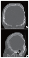
The scalp is the thickest skin in the body and protects the intracranial structures. The coverage of a large scalp defect is a difficult surgical procedure, the full details of which must be considered prior to the procedure, such as defect size and depth, and various factors related to the patient's general condition. Although a free flap is the recommended surgical procedure to cover large scalp defects, it is a high-risk operation that is not appropriate for all patients. As such, other surgical options must be explored. We present the case of a patient with an ulcer on the scalp after wide excision and split-thickness skin graft for squamous cell cancer. We successfully performed a reverse temporalis muscle flap for this patient.
Keywords: Case reports; Reconstructive surgical procedures; Scalp; Ulcer.
Conflict of interest statement
No potential conflict of interest relevant to this article was reported.
Figures


Similar articles
-
Reconstruction of a scalp defect due to cochlear implant device extrusion using a temporoparietal fascia flap and a split-thickness skin graft from the scalp.Arch Craniofac Surg. 2019 Oct;20(5):319-323. doi: 10.7181/acfs.2019.00353. Epub 2019 Oct 20. Arch Craniofac Surg. 2019. PMID: 31658797 Free PMC article.
-
Single-stage full-thickness scalp reconstruction using acellular dermal matrix and skin graft.Eplasty. 2011 Jan 25;11:e4. Eplasty. 2011. PMID: 21326624 Free PMC article.
-
The use of an omental flap for the reconstruction of a burn injury to the scalp: A case report.Int J Surg Case Rep. 2018;53:420-423. doi: 10.1016/j.ijscr.2018.11.038. Epub 2018 Nov 22. Int J Surg Case Rep. 2018. PMID: 30567059 Free PMC article.
-
Large Scalp Defect Reconstruction With Tissue Expansion, Orticochea Flap, and Acellular Dermal Matrix for Soft Tissue Augmentation: A Case Report.Cureus. 2022 Aug 5;14(8):e27723. doi: 10.7759/cureus.27723. eCollection 2022 Aug. Cureus. 2022. PMID: 36106243 Free PMC article.
-
An oculoplastic use for the temporalis muscle flap.Orbit. 2009;28(5):281-4. doi: 10.3109/01676830902856260. Orbit. 2009. PMID: 19874120 Review.
Cited by
-
Use of the frontal branch of the superficial temporal artery and the postauricular vein to overcome anatomic variations of superficial temporal vessels in scalp reconstruction with free tissue transfer: a case report.Arch Craniofac Surg. 2024 Jun;25(3):145-149. doi: 10.7181/acfs.2024.00073. Epub 2024 Jun 20. Arch Craniofac Surg. 2024. PMID: 38977399 Free PMC article.
-
Reconstruction of a temporal scalp defect without ipsilateral donor vessel possibilities using a local transposition flap and a latissimus dorsi free flap anastomosed to the contralateral side: a case report.Arch Craniofac Surg. 2023 Jun;24(3):129-132. doi: 10.7181/acfs.2023.00129. Epub 2023 Jun 20. Arch Craniofac Surg. 2023. PMID: 37415470 Free PMC article.
-
Solitary fibrous tumor in the temporalis muscle: a case report and literature review.Arch Craniofac Surg. 2023 Oct;24(5):230-235. doi: 10.7181/acfs.2023.00199. Epub 2023 Oct 20. Arch Craniofac Surg. 2023. PMID: 37919910 Free PMC article.
-
Hair Transplantation on the Baldness Region with Free Latissimus Dorsi Flap for Scalp Reconstruction: A Case Report.Arch Plast Surg. 2024 Jun 10;52(1):36-40. doi: 10.1055/s-0044-1787186. eCollection 2025 Jan. Arch Plast Surg. 2024. PMID: 39845475 Free PMC article.
-
Successful replantation of an avulsed frontal scalp through microvascular anastomoses of only one artery and one vein: a case report.Arch Craniofac Surg. 2024 Apr;25(2):95-98. doi: 10.7181/acfs.2023.00122. Epub 2024 Apr 20. Arch Craniofac Surg. 2024. PMID: 38742337 Free PMC article.
References
-
- Cheung LK. The vascular anatomy of the human temporalis muscle: implications for surgical splitting techniques. Int J Oral Maxillofac Surg. 1996;25:414–21. - PubMed
-
- Chen CT, Robinson JB, Jr, Rohrich RJ, Ansari M. The blood supply of the reverse temporalis muscle flap: anatomic study and clinical implications. Plast Reconstr Surg. 1999;103:1181–8. - PubMed
Publication types
LinkOut - more resources
Full Text Sources
Research Materials

