. 2022 Oct;1516(1):162-174.
doi: 10.1111/nyas.14856. Epub 2022 Jul 10.

Computational study of ion permeation through claudin-4 paracellular channels

Affiliations

Computational study of ion permeation through claudin-4 paracellular channels

Alessandro Berselli et al. Ann N Y Acad Sci. 2022 Oct.

Abstract

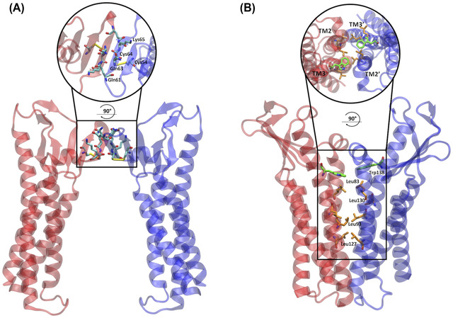
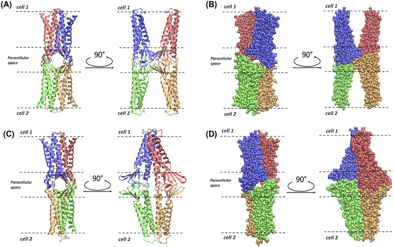
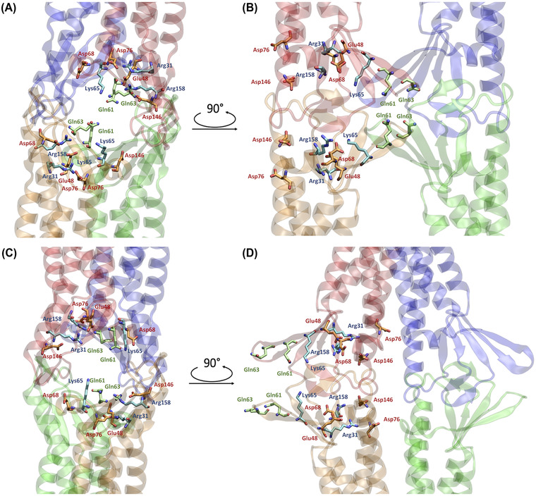
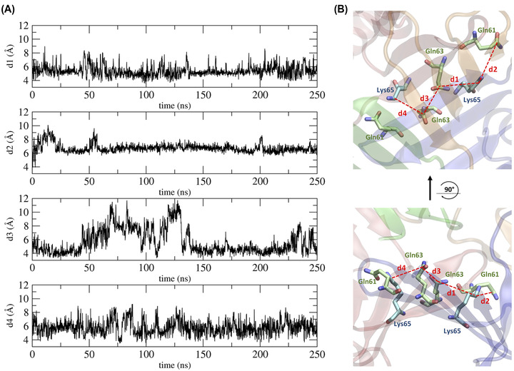
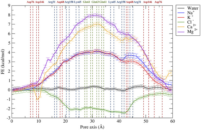
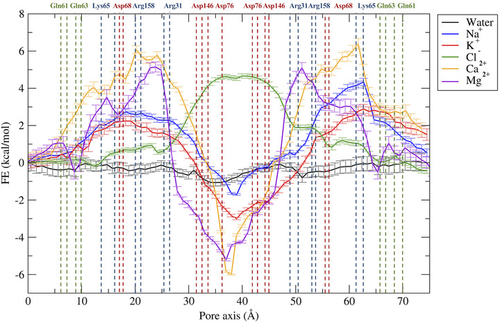
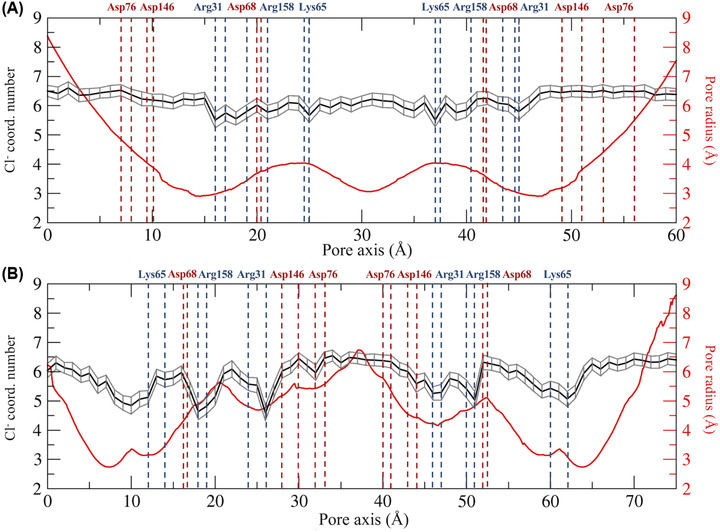
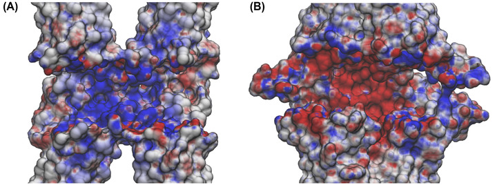
Claudins (Cldns) form a large family of protein homologs that are essential for the assembly of paracellular tight junctions (TJs), where they form channels or barriers with tissue-specific selectivity for permeants. In contrast to several family members whose physiological role has been identified, the function of claudin 4 (Cldn4) remains elusive, despite experimental evidence suggesting that it can form anion-selective TJ channels in the renal epithelium. Computational approaches have recently been employed to elucidate the molecular basis of Cldns' function, and hence could help in clarifying the role of Cldn4. In this work, we use structural modeling and all-atom molecular dynamics simulations to transfer two previously introduced structural models of Cldn-based paracellular complexes to Cldn4 to reproduce a paracellular anion channel. Free energy calculations for ionic transport through the pores allow us to establish the thermodynamic properties driving the ion-selectivity of the structures. While one model shows a cavity permeable to chloride and repulsive to cations, the other forms barrier to the passage of all the major physiological ions. Furthermore, our results confirm the charge selectivity role of the residue Lys65 in the first extracellular loop of the protein, rationalizing Cldn4 control of paracellular permeability.

Keywords: claudins; free energy calculations; ion-selectivity; molecular dynamics; paracellular space; tight junctions.


Conflict of interest statement

The authors declare no competing interests.

Figures

FIGURE 1
FIGURE 1
Dimeric cis interfaces of the two pore models. (A) In the Pore I model, two protomers of the same cell interact at the level of their ECLs, resulting in a hydrophilic interface. The apical zoomed view of the interface is shown in the circle. (B) In the Pore II model, two protomers of the same cell interact at the level of the TM domain forming a hydrophobic interface made by a leucine zipper involving the Leu83, Leu130, Leu93, and Leu127 residues and supported by the π–π interactions between the aromatic Trp138 sidechains. The apical view of the interface is shown in the circle.
FIGURE 2
FIGURE 2
Representations of the Pore I and Pore II models. (A) Apical/basolateral and lateral view of the ribbon representation of the Pore I structure. (B) Apical/basolateral and lateral view of the Van der Waals representation of Pore I. (C) Apical/basolateral and lateral view of the ribbon representation of the Pore II structure. (D) Apical/basolateral and lateral view of the Van der Waals representation of Pore II. The four Cldn4 protomers are distinguished by their coloring. In all the panels, the dashed lines identify the boundaries of the membranes of two opposing cells separated by the paracellular space.
FIGURE 3
FIGURE 3
Representations of relevant residues in the two pore models.  (A,B) Apical/basolateral A and lateral, B, views of the Pore I configuration. The β‐barrel arranged by the ECLs of the four protomers is visible. (C,D) Apical/basolateral, C, and lateral, D, views of the Pore II configuration. The reverse orientation of the pore‐lining residue compared to the Pore I is visible. The pore‐lining residues are indicated for two opposing protomers with respect to the paracellular plane. Acidic residues are depicted in orange, basic residues in light blue, and neutral residues in green. Oxygen and nitrogen atoms are shown in red and blue, respectively.
FIGURE 4
FIGURE 4
Distances between residues in the central region of the Pore I model. (A) Time evolution of distances between the amide C‐atom of the Gln residues and the amino C‐atom of the Lys residues. (B) 3D representation of the computed distances.
FIGURE 5
FIGURE 5
Free energy profiles for the permeation of water and ions through the Pore I model. The position of pore‐lining residues along the pore axis coordinates is indicated as dashed vertical lines. Acidic residues are colored in red, basic residues in blue, and neutral residues in green.
FIGURE 6
FIGURE 6
Free energy profiles for the permeation of water and ions through the Pore II model. The position of pore‐lining residues along the pore axis coordinate is indicated as dashed vertical lines. Acidic residues are colored in red, basic residues in blue, and neutral residues in green.
FIGURE 7
FIGURE 7
Hydration patterns of chloride and pore radius profile for the Pore I (A) and the Pore II (B) models. The position of the pore‐lining residues driving the ion selectivity is indicated. Acidic residues are shown in red and basic residues in blue.
FIGURE 8
FIGURE 8
Electrostatic surface of the claudin 4 tetrameric models. Cross sections of the Pore I (A) and Pore II (B) electrostatic surfaces in the paracellular region computed with the adaptive Poisson–Boltzmann solver (APBS). Potentials are shown with a red‐white‐blue color map with values ranging from −5 to +5 kT/e.

References

    1. Furuse, M. , Sasaki, H. , Fujimoto, K. , & Tsukita, S. (1998). A single gene product, claudin‐1 or ‐2, reconstitutes tight junction strands and recruits occludin in fibroblasts. Journal of Cell Biology, 143, 391–401. PMID: 9786950. - PMC - PubMed
    1. Morita, K. , Sasaki, H. , Furuse, M. , & Tsukita, S. (1999). Endothelial claudin. Journal of Cell Biology, 147, 185–194. - PMC - PubMed
    1. Anderson, J. M. (2001). Molecular structure of tight junctions and their role in epithelial transport. News in Physiological Sciences, 16, 126–130. - PubMed
    1. Farquhar, M. G. , & Palade, G. E. (1963). Junctional complexes in various epithelia. Journal of Cell Biology, 17, 375–412. PMID: 13944428. - PMC - PubMed
    1. Gumbiner, B. (1987). Structure, biochemistry, and assembly of epithelial tight junctions. American Journal of Physiology‐Cell Physiology, 253, C749–C758. - PubMed

Publication types