

Ultrasound characteristics of carotid web
- PMID: 35811446
- PMCID: PMC9544047
- DOI: 10.1111/jon.13022
Ultrasound characteristics of carotid web
Abstract
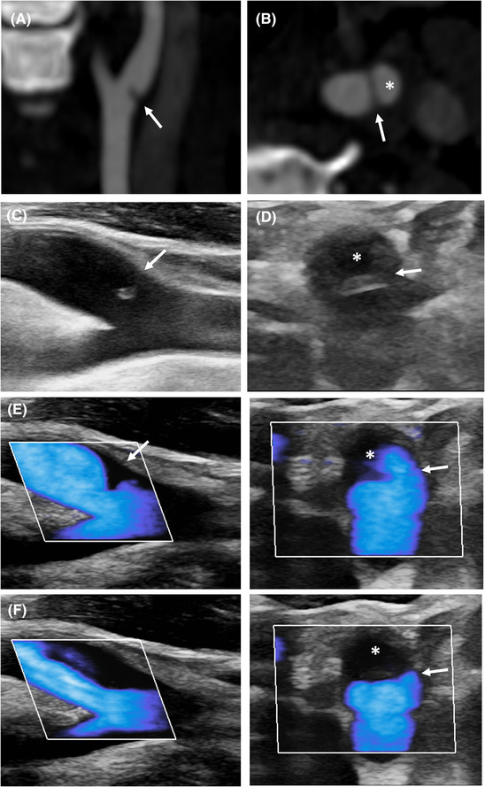
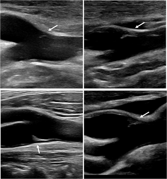
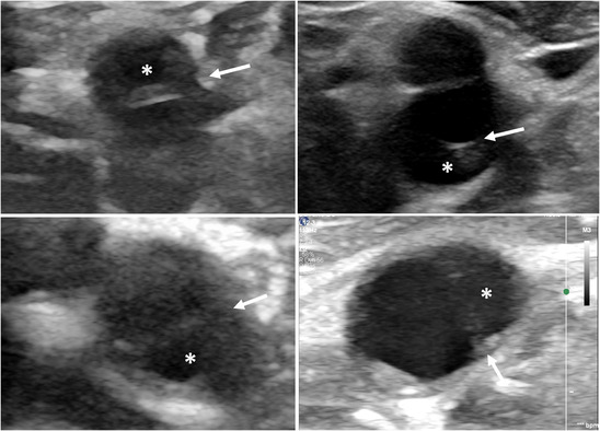
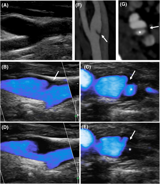
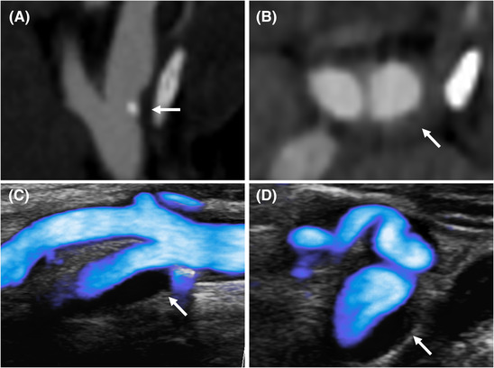
Background and purpose: Carotid web (CaW) is a cause of recurrent ischemic stroke that remains underdiagnosed using Duplex ultrasound (DUS). Improved methods and description of its ultrasound's features could allow better detection of CaW. Ultrasound microflow imaging (MFI) is a blood flow imaging technique sensitive to slow flow that could increase CaW detection. This study aimed to describe ultrasound features of CaW using B-mode imaging and MFI.
Methods: In a retrospective monocentric study, patients with CaW on CT angiography who underwent DUS examination of carotid arteries were included. DUS was performed by two nonblinded experienced neurosonologists. The specificity of CaW ultrasound features was evaluated using a group of patients with carotid atherosclerotic plaque (AP).
Results: Twenty-four patients with CaW were included. Mean age (standard deviation) was 48 years (11). Seventeen (71%) were females. Fifteen (63%) CaWs were symptomatic. MFI was available for 22 patients. B-mode imaging demonstrated the characteristic CaW appearance in 19/24 (79%) patients as a protruding triangular iso-hypoechoic lesion on longitudinal view. CaW were detected on axial view in only 9/24 (38%) patients. MFI displayed slow blood flow above CaW during systole and allowed it delineation, appearing as a thin triangular endoluminal defect in 18/22 (82%) cases. Based on MFI and B-mode, 21/22 (95%) CaWs were visible, including three CaWs only with MFI. These ultrasound features were not found among 24 patients with AP.
Conclusion: We report the ultrasound features from a series of 24 CaW. The use of MFI in addition to B-mode imaging improved the detection rate of CaW.
Keywords: Doppler ultrasound; carotid web; microflow imaging.
© 2021 The Authors. Journal of Neuroimaging published by Wiley Periodicals LLC on behalf of American Society of Neuroimaging.
Figures
References
-
- Joux J, Chausson N, Jeannin S, et al. Carotid‐bulb atypical fibromuscular dysplasia in young Afro‐Caribbean patients with stroke. Stroke 2014;45:3711‐3. - PubMed
-
- Zhang AJ, Dhruv P, Choi P, et al. A systematic literature review of patients with carotid web and acute ischemic stroke. Stroke 2018;49:2872‐6. - PubMed
MeSH terms
LinkOut - more resources
Full Text Sources
