

Efficient embryoid-based method to improve generation of optic vesicles from human induced pluripotent stem cells
- PMID: 35811797
- PMCID: PMC9218590
- DOI: 10.12688/f1000research.108829.1
Efficient embryoid-based method to improve generation of optic vesicles from human induced pluripotent stem cells
Abstract
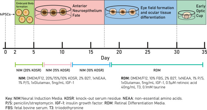
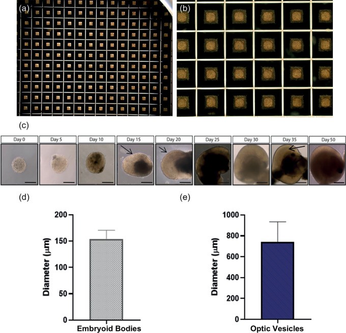
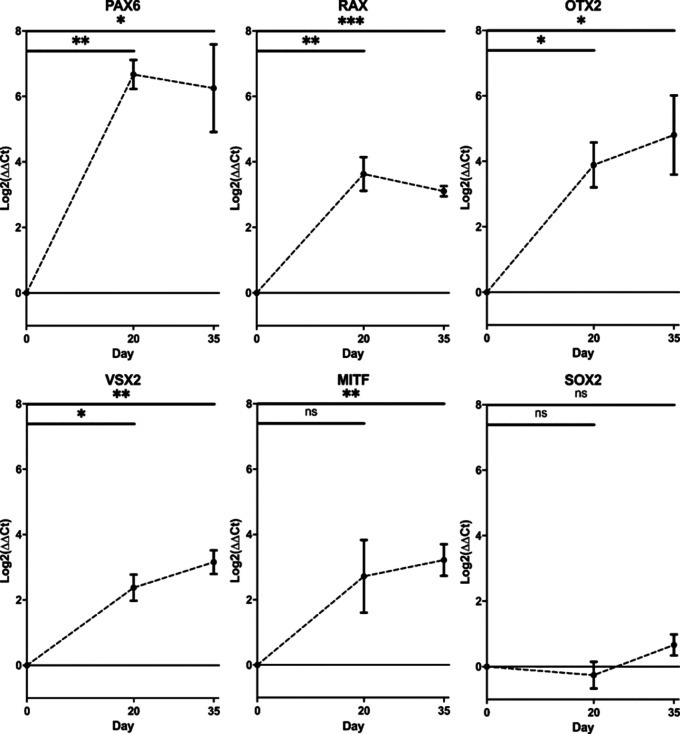
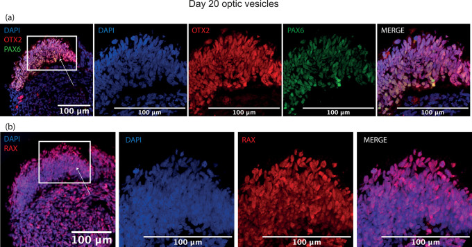
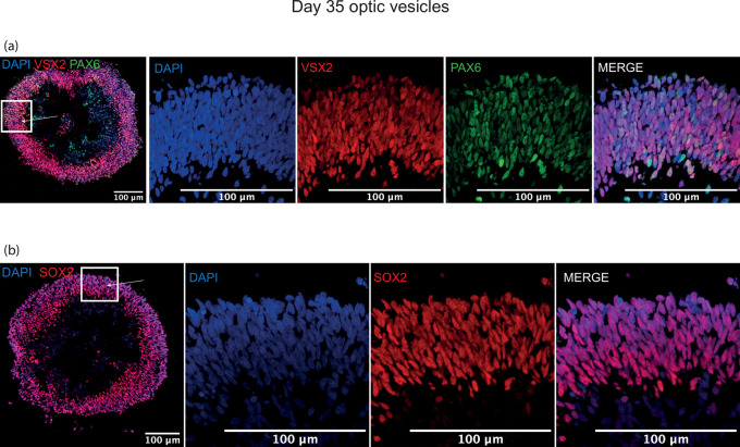
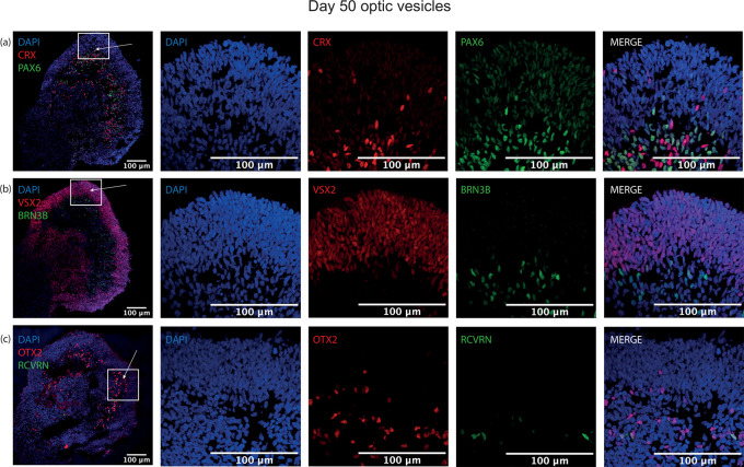
Animal models have provided many insights into ocular development and disease, but they remain suboptimal for understanding human oculogenesis. Eye development requires spatiotemporal gene expression patterns and disease phenotypes can differ significantly between humans and animal models, with patient-associated mutations causing embryonic lethality reported in some animal models. The emergence of human induced pluripotent stem cell (hiPSC) technology has provided a new resource for dissecting the complex nature of early eye morphogenesis through the generation of three-dimensional (3D) cellular models. By using patient-specific hiPSCs to generate in vitro optic vesicle-like models, we can enhance the understanding of early developmental eye disorders and provide a pre-clinical platform for disease modelling and therapeutics testing. A major challenge of in vitro optic vesicle generation is the low efficiency of differentiation in 3D cultures. To address this, we adapted a previously published protocol of retinal organoid differentiation to improve embryoid body formation using a microwell plate. Established morphology, upregulated transcript levels of known early eye-field transcription factors and protein expression of standard retinal progenitor markers confirmed the optic vesicle/presumptive optic cup identity of in vitro models between day 20 and 50 of culture. This adapted protocol is relevant to researchers seeking a physiologically relevant model of early human ocular development and disease with a view to replacing animal models.
Keywords: Embryoid bodies; PAX6; VSX2; eye development; iPSCs; optic vesicles; retinal differentiation.
Copyright: © 2022 Eintracht J et al.
Conflict of interest statement
No competing interests were disclosed.
Figures
Similar articles
-
Regulation of WNT Signaling by VSX2 During Optic Vesicle Patterning in Human Induced Pluripotent Stem Cells.Stem Cells. 2016 Nov;34(11):2625-2634. doi: 10.1002/stem.2414. Epub 2016 Jul 5. Stem Cells. 2016. PMID: 27301076 Free PMC article.
-
Modeling human retinal development with patient-specific induced pluripotent stem cells reveals multiple roles for visual system homeobox 2.Stem Cells. 2014 Jun;32(6):1480-92. doi: 10.1002/stem.1667. Stem Cells. 2014. PMID: 24532057 Free PMC article.
-
Systematic Comparison of Retinal Organoid Differentiation from Human Pluripotent Stem Cells Reveals Stage Specific, Cell Line, and Methodological Differences.Stem Cells Transl Med. 2019 Jul;8(7):694-706. doi: 10.1002/sctm.18-0267. Epub 2019 Mar 27. Stem Cells Transl Med. 2019. PMID: 30916455 Free PMC article.
-
Cell fate decisions, transcription factors and signaling during early retinal development.Prog Retin Eye Res. 2022 Nov;91:101093. doi: 10.1016/j.preteyeres.2022.101093. Epub 2022 Jul 8. Prog Retin Eye Res. 2022. PMID: 35817658 Free PMC article. Review.
-
Pluripotent Stem Cells to Model Degenerative Retinal Diseases: The RPE Perspective.Adv Exp Med Biol. 2019;1186:1-31. doi: 10.1007/978-3-030-28471-8_1. Adv Exp Med Biol. 2019. PMID: 31654384 Review.
Cited by
-
Disruption of common ocular developmental pathways in patient-derived optic vesicle models of microphthalmia.Stem Cell Reports. 2024 Jun 11;19(6):839-858. doi: 10.1016/j.stemcr.2024.05.001. Epub 2024 May 30. Stem Cell Reports. 2024. PMID: 38821055 Free PMC article.
-
Restoration of functional PAX6 in aniridia patient iPSC-derived ocular tissue models using repurposed nonsense suppression drugs.Mol Ther Nucleic Acids. 2023 Jun 26;33:240-253. doi: 10.1016/j.omtn.2023.06.016. eCollection 2023 Sep 12. Mol Ther Nucleic Acids. 2023. PMID: 37483273 Free PMC article.
-
A highly reproducible and efficient method for retinal organoid differentiation from human pluripotent stem cells.Proc Natl Acad Sci U S A. 2024 Jun 18;121(25):e2317285121. doi: 10.1073/pnas.2317285121. Epub 2024 Jun 13. Proc Natl Acad Sci U S A. 2024. PMID: 38870053 Free PMC article.
-
Robotics-Driven Manufacturing of Cartilaginous Microtissues for Skeletal Tissue Engineering Applications.Stem Cells Transl Med. 2024 Mar 15;13(3):278-292. doi: 10.1093/stcltm/szad091. Stem Cells Transl Med. 2024. PMID: 38217535 Free PMC article.
References
Publication types
MeSH terms
Substances
Grants and funding
LinkOut - more resources
Full Text Sources
Research Materials