. 2022 Jun;30(6):863-873.
doi: 10.1016/j.jsps.2022.03.005. Epub 2022 Mar 16.

Empagliflozin attenuates neurodegeneration through antioxidant, anti-inflammatory, and modulation of α-synuclein and Parkin levels in rotenone-induced Parkinson's disease in rats

Affiliations

Empagliflozin attenuates neurodegeneration through antioxidant, anti-inflammatory, and modulation of α-synuclein and Parkin levels in rotenone-induced Parkinson's disease in rats

Sanaa Ahmed et al. Saudi Pharm J. 2022 Jun.

Abstract

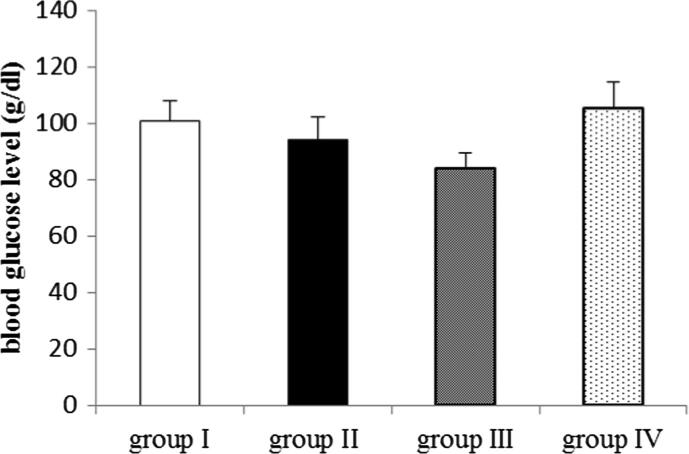
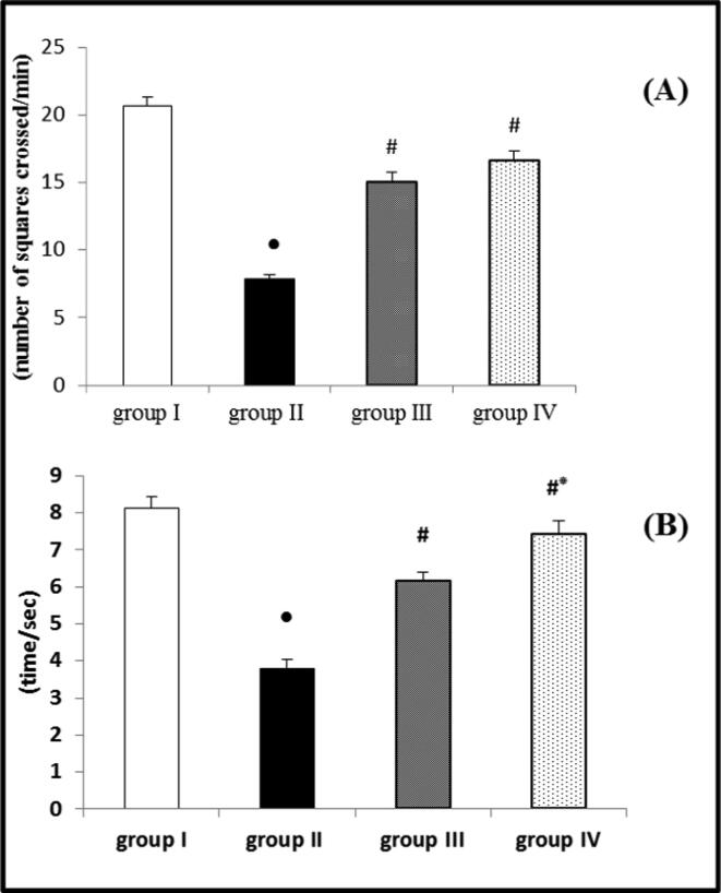
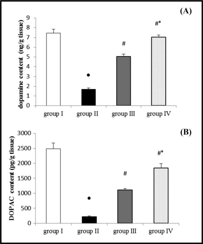
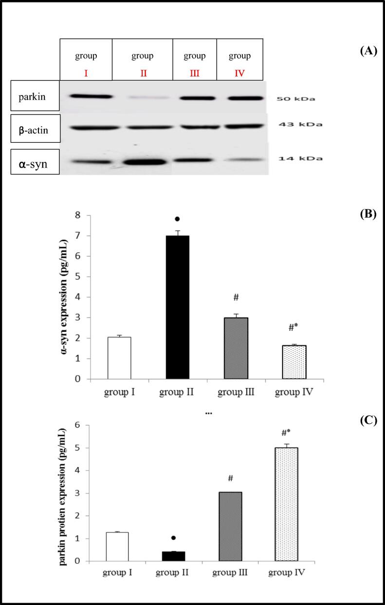
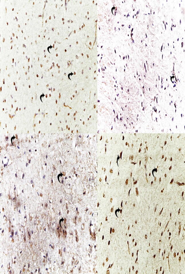
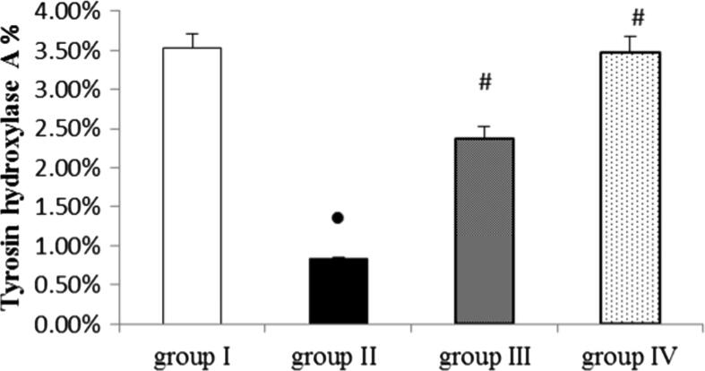
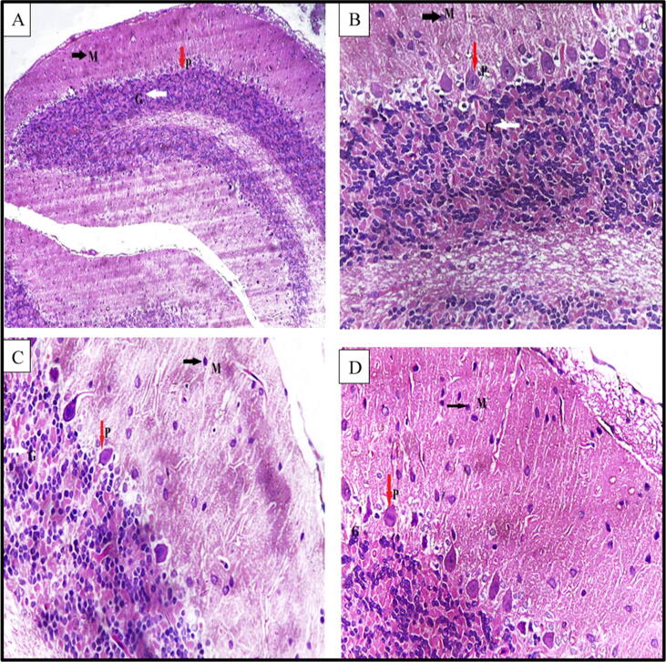
Sodium-glucose co-transporter 2 (SGLT 2) inhibitors are a relatively new antidiabetic drug with antioxidant and anti-inflammatory properties. Therefore, this study aimed to investigate whether SGLT 2 inhibitors have a neuroprotective effect in PD. Twenty-four Wistar rats were randomized into four groups. The first one (control group) received dimethyl sulfoxide (DMSO) as a vehicle (0.2 mL/48 hr, S.C). The second group (positive control) received rotenone (ROT) (2.5 mg/kg/48 hr, S.C) for 20 successive days, whereas the third and fourth groups received empagliflozin (EMP) (1 and 2 mg/kg/day, orally), respectively. The two groups received rotenone (2.5 mg/kg/48 hr S.C) concomitantly with EMP for another 20 days on the fifth day. By the end of the experimental period, behavioral examinations were done. Subsequently, rats were sacrificed, blood samples and brain tissues were collected for analysis. ROT significantly elevated oxidative stress and proinflammatory markers as well as α-synuclein. However, dopamine (DP), antioxidants, tyrosine hydroxylase (TH), and Parkin were significantly decreased. Groups of (EMP + ROT) significantly maintained oxidative stress and inflammatory markers elevation, maintained α-synuclein and Parkin levels, and elevated TH activity and dopamine level. In both low and high doses, EMP produced a neuroprotective effect against the PD rat model, with the high dose inducing a more significant effect.

Keywords: CP, Ceruloplasmin; DMSO, Dimethyl sulphoxide; DOPAC, Dihydrophenyl acetic acid; DOPAL, Dihydroxyphenylacetaldehyde; DP, Dopamine; EMP, Empagliflozin; GABA, γ-Aminobutyric acid; GSH, Reduced glutathione; IL-1β, Interlukine 1β; MAO, Monoamine oxidase; MDA, Malondialdehyde; NO, Nitric oxide; Neurodegeneration; Neuroprotection; PD, Parkinson’s disease; Parkinsonism; ROS, Reactive oxygen species; ROT, Rotenone; Rotenone; SGLT 2, Sodium glucose co-transporter 2; SGLT-2 inhibitors; SOD, Superoxide dismutase; TH, Tyrosine hydroxylase; TNF-α, Tumor necrosis factor–α; α-synuclein; α–syn, Alpha-synuclien.


Conflict of interest statement

The authors declare that they have no known competing financial interests or personal relationships that could have appeared to influence the work reported in this paper.

Figures

Fig. 1
Fig. 1
Effect of EMP on blood glucose level in ROT-induced PD in male Wistar rats. Data were presented as mean ± SEM n = 6. group I = control group. group II = ROT group. group III = EMP1mg + ROT group. group IV = EMP2mg + ROT group.
Fig. 2
Fig. 2
Effect of EMP on (A) locomotor activity determined by open field test and (B) motor coordination determined by rotarod test in ROT-induced PD in male Wistar rats. Data were presented as mean ± SEM n = 6. ● Significantly different when compared to the control group at p < 0.05. # Significantly different when compared to ROT group at p < 0.05. * Significantly different when compared to EMP (low dose) group at p < 0.05. group I = control group. group II = ROT group. group III = EMP1mg + ROT group. group IV = EMP2mg + ROT group.
Fig. 3
Fig. 3
Effect of EMP on the brain content of (A) dopamine and (B) DOPAC in ROT-induced PD in male Wistar rats. Data were presented as mean ± SEM n = 6. ● Significantly different when compared to the control group at p < 0.05. # Significantly different when compared to ROT group at p < 0.05. * Significantly different when compared to EMP (low dose) group at p < 0.05. group I = control group. group II = ROT group. group III = EMP1mg + ROT group. group IV = EMP2mg + ROT group. DOPAC = dihydrophenyl acetic acid.
Fig. 4
Fig. 4
Effect of EMP on α-syn and parkin protein expression in ROT-induced PD in male Wistar rats. Western blot bands (A), Densitometric quantitation of α-syn (B) and parkin (C) protein expressions. Data were presented as mean ± SEM n = 6. ● Significantly different when compared to the control group at p < 0.05. # Significantly different when compared to ROT group at p < 0.05. * Significantly different when compared to EMP (low dose) group at p < 0.05. group I = control group. group II = ROT group. group III = EMP1mg + ROT group. group IV = EMP2mg + ROT group. EMP = empagliflozin ROT = rotenone α-syn = α-synuclien.
Fig. 5
Fig. 5
(A) Immunohistochemical staining of striatal TH expression of the control group (400 X) showing the positive immune reaction of TH. (B): the ROT group revealed almost negligible immune reaction compared to the control group. (C and D): EMP treated groups showed restoration of staining of TH compared to the ROT group with a dose-dependent effect.
Fig. 6
Fig. 6
Quantitative image analysis for TH immunohistochemical staining presented as mean area% (A%). Data were presented as mean ± SEM n = 6. ● Significantly different when compared to the control group at p < 0.05. # Significantly different when compared to ROT group at p < 0.05. * Significantly different when compared to EMP (low dose) group at p < 0.05. group I = control group. group II = ROT group. group III = EMP1mg + ROT group. group IV = EMP2mg + ROT group.
Fig. 7
Fig. 7
A photomicrograph of a section in the cerebellar cortex of a rat (A): (control group) showing; molecular layer (M) with few scattered cells (black arrow), Purkinje cell layer (P), Purkinje cells appear as large pyriform cells arranged in a single row (red arrows) and granular cell layer (G) appears as tightly packed small cells with deeply stained nuclei (white arrow). (B): ROT group showing; molecular layer (M) few scattered cells (black arrow), Purkinje cell layer (P) with deeply stained Purkinje cells with pyknotic nuclei surrounded with vacuolated neuropil (red arrows), and granular layer (G) having cells with dense nuclei white arrow. (C): EMP1mg + ROT group showing; molecular layer (M) few scattered cells (black arrow), Purkinje cell layer (P) few numbers of Purkinje cells with dilated perineural space (red arrows), and granular layer (G) having cells with dense nuclei white arrow. (D): EMP2mg + ROT group showing nearly normal molecular layer (M) with few scattered cells (black arrow), Purkinje cell layer (P), Purkinje cells appear as large pyriform cells arranged in a single row (red arrows) and granular cell layer (G) appears as tightly packed small cells with deeply stained nuclei (white arrow). ROT = Rotenone. EMP1 = Empagliflozin 1 mg. EMP2 = Empagliflozin 2 mg. TH = tyrosine hydroxylase.

References

    1. Abdelhamid A.M., Elsheakh A.R., Abdelaziz R.R., Suddek G.M. Empagliflozin ameliorates ethanol-induced liver injury by modulating NF-κB/Nrf-2/PPAR-γ interplay in mice. Life Sci. 2020;256:117908. doi: 10.1016/j.lfs.2020.117908. - DOI - PubMed
    1. Abdelsalam R.M., Safar M.M. Neuroprotective effects of vildagliptin in rat rotenone Parkinson's disease model: role of RAGE-NF κB and Nrf2-antioxidant signaling pathways. J. Neurochem. 2015;133(5):700–707. doi: 10.1111/jnc.13087. - DOI - PubMed
    1. Acar N., Parlak H., Ozkan A., Soylu H., Avcı S., Ustunel I., Izgut-Uysal V.N., Agar A. The effect of docosahexaenoic acid on apelin distribution of nervous system in the experimental mouse model of Parkinson’s disease. Tissue Cell. 2019;56:41–51. doi: 10.1016/j.tice.2018.12.002. - DOI - PubMed
    1. Ahmed S.A., Mohammed W.I. Carvedilol induces the antiapoptotic proteins Nrf2 and Bcl2 and inhibits cellular apoptosis in aluminum-induced testicular toxicity in male Wistar rats. Biomed. Pharmacother. 2021;139:111594. doi: 10.1016/j.biopha.2021.111594. - DOI - PubMed
    1. Alabi A.O., Ajayi A.M., Ben-Azu B., Bakre A.G., Umukoro S. Methyl jasmonate abrogates rotenone-induced parkinsonian-like symptoms through inhibition of oxidative stress, release of pro-inflammatory cytokines, and down-regulation of immnopositive cells of NF-κB and α-synuclein expressions in mice. Neurotoxicology. 2019;74:172–183. doi: 10.1016/j.neuro.2019.07.003. - DOI - PubMed