

Fetal DNA Causes Sex-Specific Inflammation From Human Fetal Membranes
- PMID: 35812324
- PMCID: PMC9257279
- DOI: 10.3389/fphys.2022.901726
Fetal DNA Causes Sex-Specific Inflammation From Human Fetal Membranes
Abstract
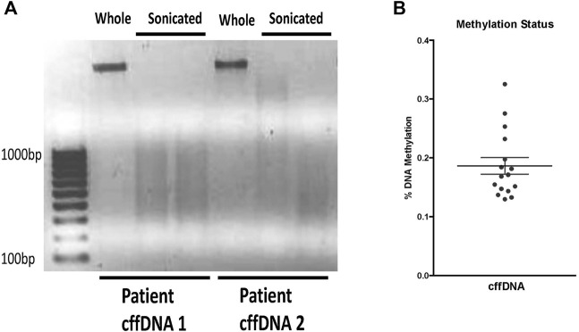
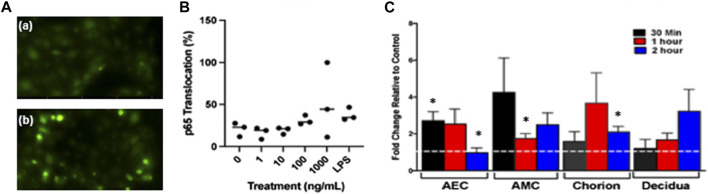
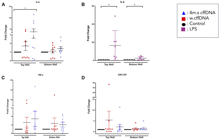
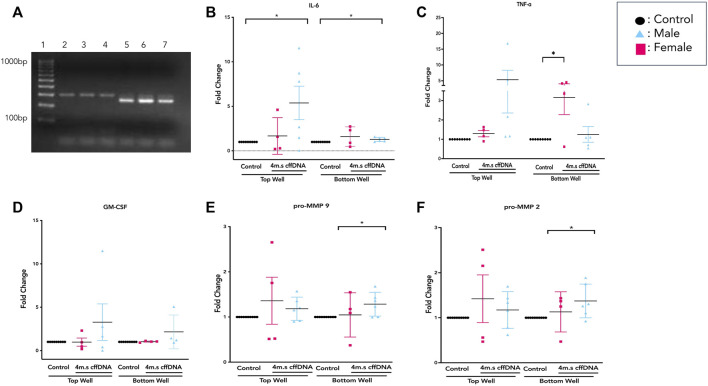
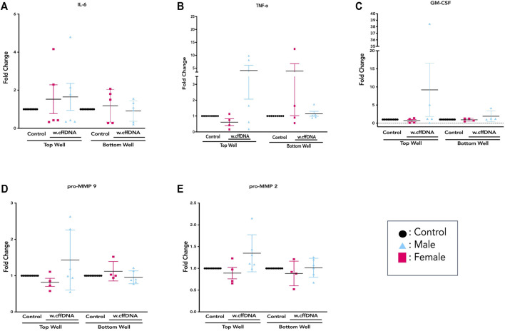
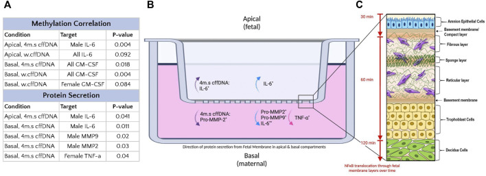
Inflammation is central to the mechanisms of parturition, but the lack of understanding of how it is controlled in normal parturition hampers our ability to understand how it may diverge resulting in preterm birth. Cell-free fetal DNA is found in the amniotic fluid, and it is thought to be able to activate inflammation as a danger-associated molecular pattern. Although its levels increases with gestational age, its effect has not been studied on the human fetal membranes. Thus, the aim of this study was to determine if the fetal DNA can trigger inflammation in the human fetal membranes and, thus, potentially contribute to the inflammatory load. Isolated human amniotic epithelial cells and fetal membrane explants were treated apically with fetal DNA causing the translocation of NF-KB into the nucleus of cells and throughout the cells of the explant layers with time. Fetal membrane explants were treated apically with either small or larger fragments of fetal DNA. IL-6, TNFα, and GM-CSF secretion was measured by ELISA, and pro-MMP2 and pro-MMP9 activity was measured by zymography from apical and basal media. Increased apical IL-6 secretion and basal pro-MMP2 activity was seen with small fragments of fetal DNA. When the data were disaggregated based on fetal sex, males had significant increases in IL-6 secretion and basal increased activity in pro-MMP2 and 9, whereas females had significantly increased basal secretion of TNFα. This was caused by the smaller fragments of fetal DNA, whereas the larger fragments did not cause any significant increases. Male fetal DNA had significantly lower percentages of methylation than females. Thus, when the cytokine and pro-MMP activity data were correlated with methylation percentage, IL-6 secretion significantly correlated negatively, whereas GM-CSF secretion positively correlated. These data support the role of fetal DNA as an inflammatory stimulus in the FM, as measured by increased NF-κB translocation, cytokine secretion, and increased pro-MMP activity. However, the data also suggested that the responses are different from FM tissues of male and female fetuses, and both the fragment size and methylation status of the fetal DNA can influence the magnitude and type of molecule secreted.
Keywords: cytokine; fetal DNA; fetal membranes; fetal sex; inflammation; methylation.
Copyright © 2022 Saito Reis, Ng, Kurashima, Padron and Kendal-Wright.
Conflict of interest statement
The authors declare that the research was conducted in the absence of any commercial or financial relationships that could be construed as a potential conflict of interest.
Figures


Similar articles
-
Stretch Causes cffDNA and HMGB1-Mediated Inflammation and Cellular Stress in Human Fetal Membranes.Int J Mol Sci. 2024 May 9;25(10):5161. doi: 10.3390/ijms25105161. Int J Mol Sci. 2024. PMID: 38791199 Free PMC article.
-
Granulocyte-macrophage colony-stimulating factor initiates amniotic membrane rupture and preterm birth in a mouse model.Am J Reprod Immunol. 2021 Aug;86(2):e13424. doi: 10.1111/aji.13424. Epub 2021 Apr 6. Am J Reprod Immunol. 2021. PMID: 33772943
-
A20, an essential component of the ubiquitin-editing protein complex, is a negative regulator of inflammation in human myometrium and foetal membranes.Mol Hum Reprod. 2017 Sep 1;23(9):628-645. doi: 10.1093/molehr/gax041. Mol Hum Reprod. 2017. PMID: 28911210
-
The Role of Danger Associated Molecular Patterns in Human Fetal Membrane Weakening.Front Physiol. 2020 Jun 17;11:602. doi: 10.3389/fphys.2020.00602. eCollection 2020. Front Physiol. 2020. PMID: 32625109 Free PMC article. Review.
-
The physiology of fetal membrane weakening and rupture: Insights gained from the determination of physical properties revisited.Placenta. 2016 Jun;42:59-73. doi: 10.1016/j.placenta.2016.03.015. Epub 2016 Apr 1. Placenta. 2016. PMID: 27238715 Review.
Cited by
-
Matrix metalloproteinases in preterm prelabor rupture of membranes in the setting of chorioamnionitis: A scoping review.Am J Reprod Immunol. 2023 Jan;89(1):e13642. doi: 10.1111/aji.13642. Epub 2022 Nov 9. Am J Reprod Immunol. 2023. PMID: 36300889 Free PMC article.
-
Stretch Causes cffDNA and HMGB1-Mediated Inflammation and Cellular Stress in Human Fetal Membranes.Int J Mol Sci. 2024 May 9;25(10):5161. doi: 10.3390/ijms25105161. Int J Mol Sci. 2024. PMID: 38791199 Free PMC article.
References
-
- Astern J. M., Collier A. C., Kendal-Wright C. E. (2013). Pre-B Cell Colony Enhancing Factor (PBEF/NAMPT/Visfatin) and Vascular Endothelial Growth Factor (VEGF) Cooperate to Increase the Permeability Of?the?human Placental Amnion. Placenta 34 (1), 42–49. 10.1016/j.placenta.2012.10.008 - DOI - PMC - PubMed
Grants and funding
LinkOut - more resources
Full Text Sources
Miscellaneous