. 2022 Jul 2:31:101299.
doi: 10.1016/j.bbrep.2022.101299. eCollection 2022 Sep.

Exosome-mediated transduction of mechanical force regulates prostate cancer migration via microRNA

Affiliations

Exosome-mediated transduction of mechanical force regulates prostate cancer migration via microRNA

Zhixiao Liu et al. Biochem Biophys Rep. .

Abstract

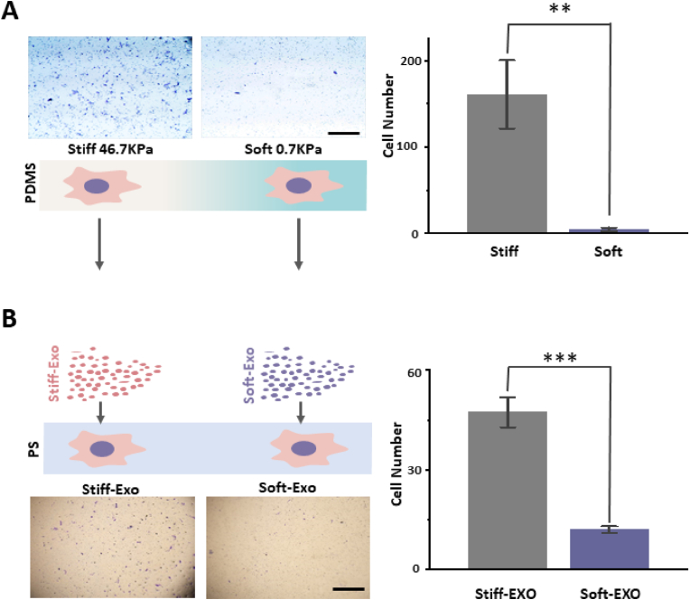
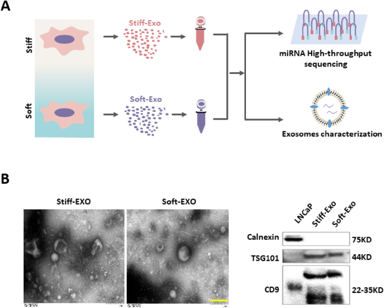
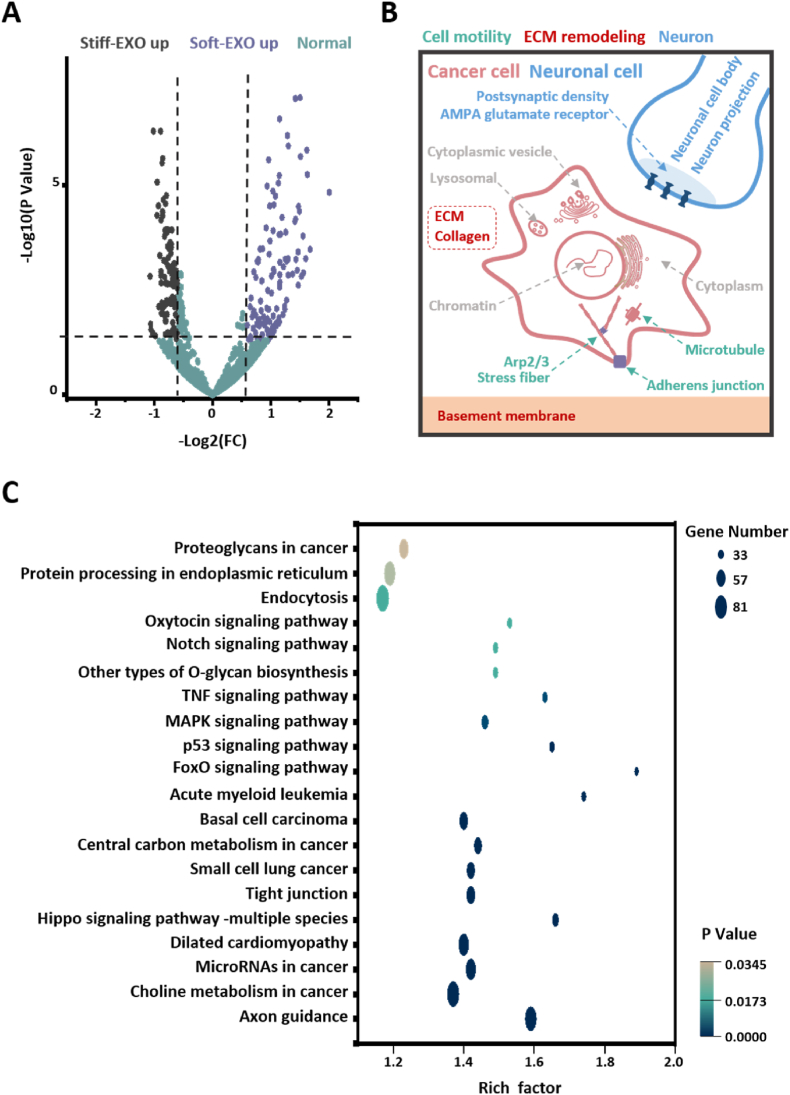
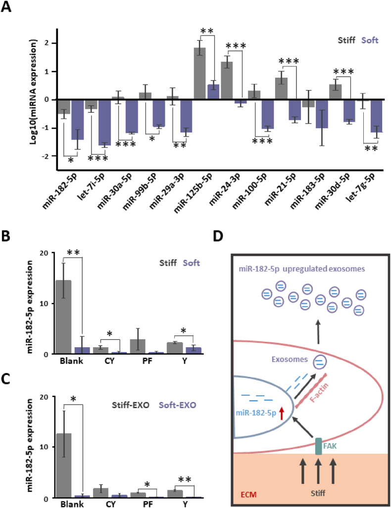
Physical cues in the extracellular microenvironment regulate cancer cell metastasis. Functional microRNA (miRNA) carried by cancer derived exosomes play a critical role in extracellular communication between cells and the extracellular microenvironment. However, little is known about the role of exosomes loaded miRNAs in the mechanical force transmission between cancer cells and extracellular microenvironment. Herein, our results suggest that stiff extracellular matrix (ECM) induced exosomes promote cancer cell migration. The ECM mechanical force regulated the exosome miRNA cargo of prostate cancer cells. Exosome miRNAs regulated by the ECM mechanical force modulated cancer cell metastasis by regulating cell motility, ECM remodeling and the interaction between cancer cells and nerves. Focal adhesion kinase mediated-ECM mechanical force regulated the intracellular miRNA expression, and F-actin mediate-ECM mechanical force regulated miRNA packaging into exosomes. The above results demonstrated that the exosome miRNA cargo promoted cancer metastasis by transmitting the ECM mechanical force. The ECM mechanical force may play multiple roles in maintaining the microenvironment of cancer metastasis through the exosome miRNA cargo.

Keywords: Cancer metastasis; Exosomes; Extracellular environment mechanical forces; MicroRNA; Prostate cancer.


Conflict of interest statement

The authors declare that they have no known competing financial interests or personal relationships that could have appeared to influence the work reported in this paper.

Figures

Fig. 1
Fig. 1
Exosomes induced by different substrate stiffnesses regulate cell migration. A. LNCaP cells were cultured on different substrates. Cell migration was analyzed after 48 h using Transwell (left, scale bar: 1 mm). LNCaP cells grown on stiff substrates had a higher migration efficiency (p = 0.00235). B. The exosomes of LNCaP cells grown on different mechanical substrates were collected and co-cultured with LNCaP grown on polystyrene (PS) petri dishes (stiff substrate-induced exosomes: Stiff-Exo; soft substrate-induced exosomes: Soft-Eox). Cell migration was analyzed after 48-h co-culture by Transwell (left, scale bar: 1 mm). LNCaP cells co-cultured with Stiff-Exo had higher migration efficiency (p = 1.87488E-4). n = 3, *p < 0.05; **p < 0.01; ***p < 0.001, t-test.
Fig. 2
Fig. 2
Collection and characterization of Stiff-Exo and Soft-Exo. A. Schematic diagram of collection and characterization of exosomes induced by different substrates stiffness. B. Characterization of Stiff-Exo and Soft-Exo morphology by transmission electron microscopy (left, bar = 200 nm). Characterization of exosome-related and cell-related protein markers by Western blot (right).
Fig. 3
Fig. 3
miRNA in LNCaP exosomes is regulated by substrate stiffness. A. Volcano map of differentially expressed miRNA in Stiff-Exo and Soft-Exo. |log2(FC)|≥0.58; p Value ≤ 0.05 is defined as difference. B. Schematic diagram of GO enrichment analysis (Cellular Component) of differential miRNAs target genes (top 19, sort according to p value). C. Enrichment analysis of differentially expressed miRNAs target genes in KEGG pathway (top 20, sort according to p value). n = 3.
Fig. 4
Fig. 4
Substrate stiffness regulates the expression of miRNA in cancer cells and transports miRNA to exosomes. A. LNCaP cells were cultured on different substrates for 48 h. Significantly different miRNA expressions in Stiff-Exo and Soft-Exo Q-PCR were detected by qRT-PCR. The results showed that, except for miR-183-5p, all other miRNAs were regulated by substrate stiffness (miR-182-5p: p = 0.0147; let-7i-5p: p = 1.24939E-4; miR-30a-5p: p = 6.96735E-4; miR-99b-5p: p = 0.0167; miR-29a-3p: p = 0.00302; miR-125b-5p: p = 0.00163; miR-24-3p: p = 6.32271E-4; miR-100-5p: p = 8.13899E-4; miR-21-5p: p = 5.60351E-4; miR-183-5p: p = 0.21238; miR-30d-5p: p = 4.79694E-4; let-7g-5p: p = 0.00562). B. LNCaP cells were cultured on different substrates for 24 h and then incubated with different inhibitors (F-actin inhibitor cytoskeleton B, CY; FAK inhibitor PF-573288, PF; ROCK inhibitor Y-27632, Y; 10 μM) for 24 h. The expression of miR-182-5p in cells was detected by qRT-PCR. The results showed that PF significantly inhibited the regulatory effect of substrate stiffness on miR-182-5p expression in cells (Blank: p = 0.00512; CY: p = 0.01741; PF: p = 0.10199; Y: p = 0.04764). C. LNCaP cells were cultured on different substrates for 24 h and then with different inhibitors for 24 h. The cell supernatant was collected to isolate Stiff-Exo and Soft-Exo. The expression of miR-182-5p in Stiff-Exo and Soft-Exo was detected by qRT-PCR. The results showed that CY significantly inhibited the regulatory effect of substrate stiffness on miR-182-5p transport to exosomes (Blank: p = 0.01; CY: p = 0.05816; PF: p = 0.01343; Y: p = 0.0031). D. Schematic diagram of the mechanism of ECM stiffness regulating miR-182-5p expression in cancer cells and transport to exosomes. The stiff ECM introduced information into LNCaP cells through FAK to promote miR-182-5p expression. LNCaP cells transferred stiff substrate-induced miR-182-5p to exosomes through F-actin. n = 3, *p < 0.05; **p < 0.01; ***p < 0.001, t-test.

References

    1. Nia H.T., Munn L.L., Jain R.K. Physical traits of cancer. Science. 2020;370 - PMC - PubMed
    1. Butcher D.T., Alliston T., Weaver V.M. A tense situation: forcing tumour progression. Nat. Rev. Cancer. 2009;9:108–122. - PMC - PubMed
    1. Liu Q., Luo Q., Ju Y., Song G. Role of the mechanical microenvironment in cancer development and progression. Cancer Biol Med. 2020;17:282–292. - PMC - PubMed
    1. Wirtz D., Konstantopoulos K., Searson P.C. The physics of cancer: the role of physical interactions and mechanical forces in metastasis. Nat. Rev. Cancer. 2011;11:512–522. - PMC - PubMed
    1. Kechagia J.Z., Ivaska J., Roca-Cusachs P. Integrins as biomechanical sensors of the microenvironment. Nat. Rev. Mol. Cell Biol. 2019;20:457–473. - PubMed

LinkOut - more resources