. 2022 Jun 22:13:873586.
doi: 10.3389/fimmu.2022.873586. eCollection 2022.

TFR Cells Express Functional CCR6 But It Is Dispensable for Their Development and Localization During Splenic Humoral Immune Responses

Affiliations

TFR Cells Express Functional CCR6 But It Is Dispensable for Their Development and Localization During Splenic Humoral Immune Responses

Cameron R Bastow et al. Front Immunol. .

Abstract

Follicular T cells including T follicular helper (TFH) and T follicular regulatory (TFR) cells are essential in supporting and regulating the quality of antibody responses that develop in the germinal centre (GC). Follicular T cell migration during the propagation of antibody responses is largely attributed to the chemokine receptor CXCR5, however CXCR5 is reportedly redundant in migratory events prior to formation of the GC, and CXCR5-deficient TFH and TFR cells are still capable of localizing to GCs. Here we comprehensively assess chemokine receptor expression by follicular T cells during a model humoral immune response in the spleen. In addition to the known follicular T cell chemokine receptors Cxcr5 and Cxcr4, we show that follicular T cells express high levels of Ccr6, Ccr2 and Cxcr3 transcripts and we identify functional expression of CCR6 protein by both TFH and TFR cells. Notably, a greater proportion of TFR cells expressed CCR6 compared to TFH cells and gating on CCR6+CXCR5hiPD-1hi T cells strongly enriched for TFR cells. Examination of Ccr6-/- mice revealed that CCR6 is not essential for development of the GC response in the spleen, and mixed bone marrow chimera experiments found no evidence for an intrinsic requirement for CCR6 in TFR cell development or localisation during splenic humoral responses. These findings point towards multiple functionally redundant chemotactic signals regulating T cell localisation in the GC.

Keywords: CCR6; T follicular helper (Tfh) cell; T follicular regulatory (Tfr) cells; chemokine receptor; germinal center T cell.


Conflict of interest statement

The authors declare that the research was conducted in the absence of any commercial or financial relationships that could be construed as a potential conflict of interest.

Figures

Figure 1
Figure 1
Chemokine receptor gene expression by follicular T cells. (A) Representative gating strategy for sorting follicular T cells (CD4+TCRβ+CXCR5hiPD-1hi) from day 5 SRBC i.p. immunized mice. Expression of (B) follicular T cell master transcription factor Bcl6, (C) known follicular T cell-tropic chemokine receptors, and (D) all other known classical chemokine receptors, between in naïve CD4 T cells (CD4+CD44-CD62L+) and follicular T cells, relative to Gapdh. (B–D) Data pooled from two independent experiments with 3 mice pooled per time point ± SEM, two-tailed unpaired Student’s t test. *p < 0.05, **p < 0.01.
Figure 2
Figure 2
CCR6 expression is highest in TFR cells amongst follicular T cell populations. (A, B) Representative gating strategy for CCR6+ TFH cells (CD4+B220-CXCR5hiPD-1hiFoxp3-), TFR cells (CD4+B220-CXCR5hiPD-1hiFoxp3+), naïve CD4 T cells (CD4+B220-CD44loFoxp3-) and natural T-regulatory cells (nTreg: CD4+B220-CD44midFoxp3+Nrp-1+) 6 days after SRBC immunization. (C) Geometrical mean fluorescence intensity (gMFI) of CCR6 and, (D) percentage of CCR6+ cells within populations from (A) and (B). (E) Representative gating strategy for TFH (Foxp3-) and TFR cells (Foxp3+) within CCR6+ follicular T cells (CD4+B220-CXCR5hiPD-1hiCCR6+). (F) Ratio of TFH : TFR cells within gating strategies from (A, E). (G) Frequency of CCR6+ TFH and TFR cells at indicated time points after i.p. NP-KLH/Alum immunization. (A–C, E, F) Data representative of two independent experiments, n=4 mice ± SEM, one-way ANOVA with Tukey’s multiple comparisons test, (G) n=4-5 mice per time-point ± SEM, two-way ANOVA with Sidak’s multiple comparison test. NS, Not significant, *p < 0.05, ***p < 0.001, ****p < 0.0001.
Figure 3
Figure 3
Follicular T cell populations migrate ex vivo to CCL20. (A–E) Transwell migration of splenocytes from day 6 SRBC immunized WT or Ccr6 -/- mice to increasing concentrations of CCL20. Cell populations were identified within total migrated splenocytes by flow cytometry and migration index was calculated relative to controls without chemokine. (A) Naïve CD4 T cells: CD4+B220-CXCR5-CD44loFoxp3-, (B) B cells: CD4-B220+, (C) nTregs: CD4+B220-CXCR5-CD44midFoxp3+Nrp-1+, (D) TFH cells: CD4+B220-CXCR5hiPD-1hiCD44hiFoxp3-, and (E) TFR cells: CD4+B220-CXCR5hiPD-1hiCD44hiFoxp3+. Data pooled from two independent experiments, n=3-5 mice/strain ± SEM, two-tailed unpaired Student’s t test between strains at each concentration. *p < 0.05, **p < 0.01.
Figure 4
Figure 4
CCR6-deficient mice mount normal TFH and TFR cell responses. (A) Representative gating strategy of TFH cells (CD4+B220-CXCR5hiPD-1hiCD44hiFoxp3-) and TFR cells (CD4+B220-CXCR5hiPD-1hiCD44hiFoxp3+) in WT and Ccr6 -/- mice four- and six-days post SRBC immunization. Frequency and number of (B) TFH and (C) TFR cells from (A). (D) Ratio of TFH : TFR cells four- and six-days post SRBC immunization in WT and Ccr6 -/- mice. (A–D) Data representative of 3 independent experiments, n=6-7 mice/time point ± SEM, two-tailed unpaired Student’s t test between strains at each time point.
Figure 5
Figure 5
CCR6-deficiency does not affect splenic Foxp3+ cell distribution during the peak of SRBC immunization. (A) Localisation of Foxp3+ cells in PFA and acetone fixed/permeabilized spleen sections from day 6 i.p. SRBC immunized WT and Ccr6 -/- mice. Sections were stained with antibodies against CD4 (red), IgD (blue) and Foxp3 (green). Based on CD4 and IgD staining, the following areas are outlined: follicles (blue), GCs (green), T-B border (magenta), and T-zone (red). Scale bar: 200µm. (B) Identification of Foxp3+ cells from (A), colour-coded based on localisation within the T-zone (red), T-B border (magenta), follicle (blue), or GC (green). (C) Average area (mm2) of delineated splenic compartments from (A). (D) Percentage and (E) number/mm2 of Foxp3+ cells within each splenic niche, quantified from Figure 5.B. (A, B) Images representative of n=5 mice/strain, 2-6 images/mouse. (C–E) Each dot represents the average of technical replicates per biological replicate, n=5 mice/strain ± SEM. Two-tailed unpaired Student’s t test.
Figure 6
Figure 6
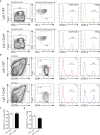
Cell-intrinsic CCR6 function is not required for the formation of TFH and TFR cell populations. Representative gating strategies of (A) naïve CD4 T cells and nTregs, and (B) TFH and TFR cells from day 6 SRBC immunized irradiated Ly5.1 hosts reconstituted with a 1:1 ratio of Ly5.1:CD45.2+ Ccr6 +/+ (WT, top row) or CD45.2+ Ccr6 -/- (bottom row) bone marrow. (C) Ratio of CD45.2+ TFH cells:CD45.2+ naïve CD4 T cells in Ly5.1:WT and Ly5.1:Ccr6 -/- chimeras. (D) Ratio of CD45.2+ TFR cells:CD45.2+ nTregs in Ly5.1:WT and Ly5.1:Ccr6 -/- chimeras. (A–D) n=6/chimera group, ± SEM, two-tailed unpaired Student’s t test.

References

    1. Mintz MA, Cyster JG. T Follicular Helper Cells in Germinal Center B Cell Selection and Lymphomagenesis. Immunol Rev (2020) 296:48–61. doi: 10.1111/imr.12860 - DOI - PMC - PubMed
    1. Deng J, Wei Y, Fonseca VR, Graca L, Yu D. T Follicular Helper Cells and T Follicular Regulatory Cells in Rheumatic Diseases. Nat Rev Rheumatol (2019) 15:475–90. doi: 10.1038/s41584-019-0254-2 - DOI - PubMed
    1. Botta D, Fuller MJ, Marquez-Lago TT, Bachus H, Bradley JE, Weinmann AS, et al. . Dynamic Regulation of T Follicular Regulatory Cell Responses by Interleukin 2 During Influenza Infection. Nat Immunol (2017) 18:1249–60. doi: 10.1038/ni.3837 - DOI - PMC - PubMed
    1. Clement RL, Daccache J, Mohammed MT, Diallo A, Blazar BR, Kuchroo VK, et al. . Follicular Regulatory T Cells Control Humoral and Allergic Immunity by Restraining Early B Cell Responses. Nat Immunol (2019) 20:1360–71. doi: 10.1038/s41590-019-0472-4 - DOI - PMC - PubMed
    1. Fu W, Liu X, Lin X, Feng H, Sun L, Li S, et al. . Deficiency in T Follicular Regulatory Cells Promotes Autoimmunity. J Exp Med (2018) 215:815–25. doi: 10.1084/jem.20170901 - DOI - PMC - PubMed

LinkOut - more resources