. 2022 Jun 23:13:917014.
doi: 10.3389/fimmu.2022.917014. eCollection 2022.

DDOST Correlated with Malignancies and Immune Microenvironment in Gliomas

Affiliations

DDOST Correlated with Malignancies and Immune Microenvironment in Gliomas

Xiaojing Chang et al. Front Immunol. .

Abstract

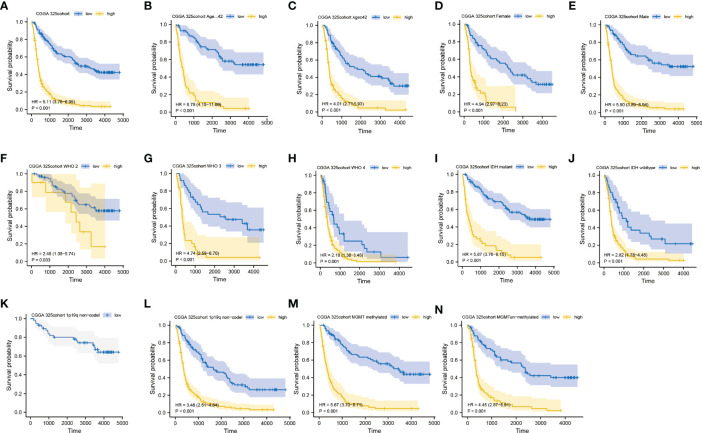
Among the most common types of brain tumor, gliomas are the most aggressive and have the poorest prognosis. Dolichyl-diphosphooligosaccharide protein glycosyltransferase non-catalytic subunit (DDOST) encodes a component of the oligosaccharide transferase complex and is related to the N-glycosylation of proteins. The role of DDOST in gliomas, however, is not yet known. First, we performed a pan cancer analysis of DDOST in the TCGA cohort. The expression of DDOST was compared between glioma and normal brain tissues in the GEO and Chinese Glioma Genome Atlas (CGGA) databases. In order to explore the role of DDOST in glioma, we analyze the impact of DDOST on the prognosis of glioma patients, with the CGGA 325 dataset as a test set and the CGGA 693 dataset as a validation set. Immunohistochemistry was performed on tissue microarrays to examine whether DDOST has an impact on glioma patient survival. Next, using single-cell sequencing analysis, GSEA, immune infiltration analysis, and mutation analysis, we explored how DDOST affected the glioma tumor microenvironment. Finally, we evaluated the clinical significance of DDOST for glioma treatment by constructing nomograms and decision curve analysis (DCA) curves. We found that DDOST was overexpressed in patients with high grade, IDH wild type, 1p19q non-codel and MGMT un-methylated, which was associated with poor prognosis. Patients with high levels of DDOST, regardless of their clinical characteristics, had a worse prognosis. Immunohistochemical analysis confirmed the results of the above bioinformatics analysis. Mechanistic analysis revealed that DDOST was closely associated with the glioma microenvironment and negatively related to tumor-infiltrating B cells and CD4+ T cells and positively related to CAFs and tumor-associated macrophages. In conclusion, these findings suggested that DDOST mediated the immunosuppressive microenvironment of gliomas and could be an important biomarker in diagnosing and treating gliomas.

Keywords: DDOST; glioma; microenvironment; prognosis; progression.


Conflict of interest statement

The authors declare that the research was conducted in the absence of any commercial or financial relationships that could be construed as a potential conflict of interest.

Figures

Figure 1
Figure 1
Pan cancer analysis of DDOST expression. (A) Analysis of DDOST expression in 33 tumors on TCGA. *, **, ***, **** indicate p < 0.05, p < 0.01, p < 0.001, and p < 0.0001, respectively; ns, not significant (Wilcoxon test). (B) Risk plot of correlation between DDOST levels and OS.
Figure 2
Figure 2
Expression difference of DDOST between glioma and normal tissue. (A) The expression of DDOST was different between glioma and normal brain tissue in the CGGA cohort. (B, C) In the GSE4290 and GSE50161 cohort, DDOST expression was significantly higher in glioma patients than in normal brain. (D, E) In the TCGA and GTEx, the expression of DDOST in LGG and GBM was significantly higher than that in normal tissue. *p < 0.05, ***p < 0.001.
Figure 3
Figure 3
Expression difference of DDOST between different clinical characters in patients with glioma in the CGGA 325 cohort. The expression of DDOST in different age (A), gender (B), PRS type (C), grade (D), IDH (E), 1p19q (F), and MGMT status (G). *p < 0.05, ***p < 0.001. ns, not significant.
Figure 4
Figure 4
Prediction of outcome of the DDOST in stratified patients in the CGGA 325 dataset. Survival curve was used to analyze OS in the low- and high-DDOST groups in CGGA 325 set (A). Survival analysis of the signature in patients stratified by age (B, C), gender (D, E), grade (F, H), IDH (I, J), 1p19q status (K, L), and MGMT promoter (M, N).
Figure 5
Figure 5
The expression of DDOST in gliomas and its prognostic significance were analyzed by immunohistochemistry. (A–C) showed that DDOST is strongly, moderately, and weakly positive in gliomas, respectively. (D) shows the expression of DDOST in normal brain tissue. High DDOST expression in glioma was related to poor OS (E).
Figure 6
Figure 6
The expression of DDOST in glioma by single-cell analysis. (A) By dimensionality reduction analysis of CGGA single-cell data, 6,148 cells were divided into astrocytes, epithelial cells, macrophages, monocytes, and T cells. (B) Scatter plot of DDOST distribution in glioma. (C) The scatter plot shows the expression of DDOST in astrocytes, epithelial cells, macrophages, monocytes, and T cells. ***p < 0.001.
Figure 7
Figure 7
GO and KEGG functional enrichment analysis of the role of DDOST in glioma. (A) The volcano map shows the differential genes between high- and low-DDOST groups by the CGGA 325 cohort (A). KEGG (B) and GO (C) were used to analyze the relevant mechanisms.
Figure 8
Figure 8
Immune infiltration patterns of low- and high-DDOST analyzed by ssGSEA methods in glioma from the CGGA dataset. (A) Heatmap revealing the scores of immune cells in low and high immunities. (B–E) Scatter plot showing the correlation between DDOST and tumor purity, stromal, ESTIMATE, and immune scores.
Figure 9
Figure 9
The relationship between the expression of DDOST and immune cells were analyzed by EPIC. The scatter plot shows the correlation between DDOST and B cells (A), CAFs (B), CD4+ T cells (C), CD8+ T cells (D), macrophages (E), NK cells (F), and other cells (G).
Figure 10
Figure 10
The relationship between DDOST and tumor immune infiltration was analyzed by NMF cluster analysis of glioma patients with immune-related genes. (A) According to the cophenetic value and the expression of immune-related genes, glioma patients were divided into two clusters, C1 and C2. (B) The survival curve was used to analyze the survival difference between C1 and C2 groups. (C) The expression differences of DDOST in C1 and C2 clusters were compared. ***p < 0.001.
Figure 11
Figure 11
Nomogram for the prediction of prognostic probabilities in the CGGA dataset. (A) The nomogram for the prediction of OS was developed using the CGGA dataset. (B) The calibration plots for predicting 1-, 3-, and 5-year survival. (C) Decision curve analysis for the DDOST nomogram and the clinicopathological nomogram to estimate the OS.

Similar articles

Cited by

References

    1. Ostrom QT, Cioffi G, Waite K, Kruchko C, Barnholtz-Sloan JS. CBTRUS Statistical Report: Primary Brain and Other Central Nervous System Tumors Diagnosed in the United States in 2014-2018. Neuro Oncol (2021) 23(12 Suppl 2):iii1–1iii105. doi: 10.1093/neuonc/noab200 - DOI - PMC - PubMed
    1. Louis DN, Perry A, Wesseling P, Brat DJ, Cree IA, Figarella-Branger D, et al. . The 2021 WHO Classification of Tumors of the Central Nervous System: A Summary. Neuro Oncol (2021) 23(8):1231–51. doi: 10.1093/neuonc/noab106 - DOI - PMC - PubMed
    1. Yang Z, Ling F, Ruan S, Hu J, Tang M, Sun X, et al. . Clinical and Prognostic Implications of 1p/19q, IDH, BRAF, MGMT Promoter, and TERT Promoter Alterations, and Expression of Ki-67 and P53 in Human Gliomas. Cancer Manag Res (2021) 13:8755–65. doi: 10.2147/CMAR.S336213 - DOI - PMC - PubMed
    1. Schjoldager KT, Narimatsu Y, Joshi HJ, Clausen H. Global View of Human Protein Glycosylation Pathways and Functions. Nat Rev Mol Cell Biol (2020) 21(12):729–49. doi: 10.1038/s41580-020-00294-x - DOI - PubMed
    1. Oliveira-Ferrer L, Legler K, Milde-Langosch K. Role of Protein Glycosylation in Cancer Metastasis. Semin Cancer Biol (2017) 44:141–52. doi: 10.1016/j.semcancer.2017.03.002 - DOI - PubMed

Publication types