. 2022 Jun 22:13:916187.
doi: 10.3389/fimmu.2022.916187. eCollection 2022.

Fibrin(ogen) Is Constitutively Expressed by Differentiated Intestinal Epithelial Cells and Mediates Wound Healing

Affiliations

Fibrin(ogen) Is Constitutively Expressed by Differentiated Intestinal Epithelial Cells and Mediates Wound Healing

Amira Seltana et al. Front Immunol. .

Abstract

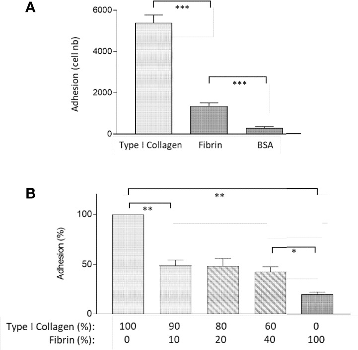
Fibrinogen is a large molecule synthesized in the liver and released in the blood. Circulating levels of fibrinogen are upregulated after bleeding or clotting events and support wound healing. In the context of an injury, thrombin activation drives conversion of fibrinogen to fibrin. Fibrin deposition contains tissue damage, stops blood loss, and prevents microbial infection. In most circumstances, fibrin needs to be removed to allow the resolution of inflammation and tissue repair, whereas failure of this may lead to the development of various disorders. However, the contribution of fibrinogen to tissue inflammation and repair is likely to be context-dependent. In this study, the concept that fibrin needs to be removed to allow tissue repair and to reduce inflammation is challenged by our observations that, in the intestine, fibrinogen is constitutively produced by a subset of intestinal epithelial cells and deposited at the basement membrane as fibrin where it serves as a substrate for wound healing under physiological conditions such as epithelial shedding at the tip of the small intestinal villus and surface epithelium of the colon as well as under pathological conditions that require rapid epithelial repair. The functional integrity of the intestine is ensured by the constant renewal of its simple epithelium. Superficial denuding of the epithelial cell layer occurs regularly and is rapidly corrected by a process called restitution that can be influenced by various soluble and insoluble factors. Epithelial cell interaction with the extracellular matrix greatly influences the healing process by acting on cell morphology, adhesion, and migration. The functional contribution of a fibrin(ogen) matrix in the intestine was studied under physiological and pathological contexts. Our results (immunofluorescence, immunoelectron microscopy, and quantitative PCR) show that fibrin(ogen) is a novel component of the basement membrane associated with the differentiated epithelial cell population in both the small intestine and colon. Fibrin(ogen) alone is a weak ligand for epithelial cells and behaves as an anti-adhesive molecule in the presence of type I collagen. Furthermore, the presence of fibrin(ogen) significantly shortens the time required to achieve closure of wounded epithelial cell monolayers and co-cultures in a PI3K-dependent manner. In human specimens with Crohn's disease, we observed a major accumulation of fibrin(ogen) throughout the tissue and at denuded sites. In mice in which fibrin formation was inhibited with dabigatran treatment, dextran sulfate sodium administration provoked a significant increase in the disease activity index and pathological features such as mucosal ulceration and crypt abscess formation. Taken together, these results suggest that fibrin(ogen) contributes to epithelial healing under both normal and pathological conditions.

Keywords: Crohn’s disease; PI3K; dabigatran; epithelial restitution; fibrinogen; human; intestinal homeostasis; mouse.


Conflict of interest statement

The authors declare that the research was conducted in the absence of any commercial or financial relationships that could be construed as a potential conflict of interest.

Figures

Figure 1
Figure 1
Epithelial FBG expression in the human intestinal mucosa. (A, B) Indirect immunofluorescence using the anti-FBG 15H12 antibody in red and α-SMA in green confirmed FBG deposition in the normal human intestine. Immunoreactive FBG was detected at the epithelial–stromal interface of the upper third regions of the villus in the adult small intestine (A) and at the base of the colonocytes of the surface epithelium of the adult colon (B). FBG was also detected in the human fetal intestine at the epithelial–stromal interface in the upper half of the villi with the anti-FBG F0111 antibody in green (tissues were counterstained with Evan blue—brown-red staining). (C) No specific anti-FBG staining was detected in the region of the crypts (* in A–C). (D) Analysis of stromal and epithelium RNA fractions confirms the epithelial origin of FBG in the human gut. RT-PCR analysis of sucrase-isomaltase (SI), an exclusive intestinal epithelial cell marker, and tenascin-C (TenC), a stromal cell marker, confirmed that the FBG beta chain (FBβ) was expressed in the epithelial fraction of the mid-gestation intestine. RPLP0 was used as a loading control. (E–G) Representative double staining illustration of FBG at the tip of the villus using the anti-FBG F0111 antibody in green (E, G) and anti-fibrin 59D8 in red (F,G) showing that a significant part of FBG at this site is under the form of fibrin. (H–J) Representative double staining illustration for the immunodetection of FBG with the anti-F0111 antibody in green (H, J) and the anti-laminin beta chain 2E8 antibody in red (I, J) at the tip of the villus showing similar deposition at the epithelial–stromal interface. Scale bars: (A, B) 100 µm and (C) 50 µm.
Figure 2
Figure 2
Ultrastructural immunolocalization of FBG at the intestinal epithelial–stromal interface. (A) Low magnification of the epithelial cell-stromal region showing positive intracellular staining at the base of the epithelial cells (e) and immunoreactive material over the extracellular components surrounding the subepithelial myofibroblasts (mf) with the exception of the epithelial basal lamina (bl) where only low-density gold staining is observed. (B) Low magnification of the apical region of an epithelial cell showing only background staining. (C) High magnification of the epithelial–stromal interface showing the lack of immunoreactive staining at the base of the epithelial cell, plasma membrane (arrows), and basal lamina (bl) while immunogold particles over the extracellular material of the pars fibroreticularis (pfr). Scales bars: (A, B) 500 nm and (C) 200 nm.
Figure 3
Figure 3
The spatial-temporal expression of FBG in intestine epithelial cells. (A) Expression of FBG in epithelial intestinal cell models that recapitulate the crypt-villus axis revealed an increase of expression of the FBG β-chain transcript in Caco-2/15 cells, closely accompanying the expression of the differentiation marker sucrase-isomaltase (SI) over the first 15 days of post-confluence (PC), whereas neither FBG nor SI was detected in HIEC-6 cells. Data are expressed as fold relative to small intestinal extracts. (B, C) When investigated at the protein level, FBG was only detected at later stages of post confluent Caco-2/15 cell culture either under its native 340-KDa form (B) or as insoluble extracellular material deposited by post confluent Caco-2/15 cells detected as 55kDa FBG fragment (C). (D–F) Immunofluorescent detection of FBG with the anti-FBG F0111 antibody (in green) on cryosections of mono- and co-cultures cells. The FBG was found to accumulate at the base (arrowheads) of post confluent Caco-2/15 monolayers (E). FBG was not detected in the multilayer of fetal stromal cells grown alone (D). In co-cultures of Caco-2/15 cells (e) on top of fetal stromal (s) cells, FBG staining was observed at the base of the epithelial cells and at the epithelial–stromal interface (arrowheads) (F). Scale bars: 25 µm.
Figure 4
Figure 4
Fibrin acts as an anti-adhesive ECM for epithelial cells. (A) Caco-2/15 showed a 70% decrease of adhesion on fibrin coating as compared to type I collagen in 30-min adhesion assay. (B) Combined with collagen I, fibrin negatively affects the adhesive properties of the collagen gel, even when added at a low concentration, in an overnight adhesion assay. At least three independent experiments were performed for each condition, and the results are expressed as means ± SEM. *p < 0.05; **p < 0.01; ***p < 0.001.
Figure 5
Figure 5
Assembly of FBG by intestinal epithelial cells and promotion of epithelial restitution on plastic and co-culture. (A, B) Caco-2/15 cells were grown on glass coverslips and then overlaid with medium alone (A) or containing plasma FBG (B) for 24 h before washing and processed for immunodetection of FBG and stained with DAPI. In comparison with control (A), cells incubated with FBG showed a fibrillar pattern of deposition at their ECM (B). Scale bars: 50 µm. (C,D) Caco-2/15 cells were grown to 12 days PC before micro-wounded and treated with hydroxyurea to evaluate the effect of extracellular components added to the medium on epithelial restitution. On plastic (C), cell migration was not accelerated by the addition of type 1 collagen or fibronectin, whereas FBG had an effect after 48 h. In co-cultures with stromal cells (B), all treatments stimulated cell migration compared to plastic assays so that wound healing was complete after 24 h for the three tested components. FBG was almost twice as efficient as collagen I or fibronectin so that 50% of the wound closure was reached in less than 4 h and more than 85% 12 h after wounding. At least three independent experiments were performed, and the results are expressed as means ± SEM. **p < 0.01; ***p < 0.001 vs. control at the same time.
Figure 6
Figure 6
FBG migration cells were in a PI3K-dependent manner. Micro-lesions were induced in 12-day PC Caco-2/15 monolayers sitting on stromal cells, followed by PI3K inhibitor (LY294002) and/or FBG treatment for 4 or 24 h. LY294002 had no significant effect on Caco-2/15 grown without FBG. In cells grown in the presence of FBG, epithelial cell restitution was inhibited by LY294002 at both 4 and 24 h. At least three independent experiments were performed, and the results are expressed as means ± SEM. ***p < 0.001.
Figure 7
Figure 7
FBG expression in Crohn’s disease. Specimens from five patients with Crohn’s disease were tested for FBG expression using the anti-FBG F0111 antibody showed in red. (A) Resection margin showing FBG deposition at the epithelial–stromal interface (arrowheads). (B) Representative low magnification of inflamed mucosa showing more intense staining of immunoreactive FBG at the epithelial–stromal interface compared to resection margin. Note that staining at the epithelial–stromal interface was stronger in the inflamed mucosa than in the resection margin at sites where the epithelium was present (C, D) or where it was denuded (B’). Additional staining was also detected throughout the lamina propria and blood vessels of the inflamed mucosa. Scale bars = 50 µm. (E) Scatter dot plot for FBG transcript analysis by qPCR in matched resection margin (RM) and inflamed mucosa (IM) paired samples from 14 patients with CD. Horizontal bars are for the medians with interquartile ranges.
Figure 8
Figure 8
Effect of thrombin inhibitor dabigatran treatment on FBG deposition in the mouse intestine and blood coagulation. Mice were treated with dabigatran for 12 days and small intestine (A, B) and colon (C, D) were harvested for control (A, C) and treated (B, D). Typical deposition of FBG (using the anti-FBG F0111) at the epithelial–stromal interface of the upper half of the villus and surface epithelium (green staining, arrowheads) as seen in the human intestine was observed in both the control mouse small intestine (A) and colon (C). Consistent reduction of staining intensity in dabigatran treated mice was noted for both small intestine (B) and colon (D) compared to controls. Tissues were counterstained with Evan blue (red staining). Bars = 100 µm.
Figure 9
Figure 9
Thrombin inhibition exacerbated DSS-induced colitis in the mouse model. Mice treated or not with dabigatran for a 12-day period received DSS in their drinking water (or regular drinking water) for the last 7 days of treatment and the effects on body weight change (A) and disease activity index (B) were monitored daily showing that thrombin inhibition predisposes mice to DSS treatment with a significant reduction of weight and increase in disease activity index. *p < 0.05 and **p < 0.01, dabigatran/DSS vs. control; ## p < 0.01, dabigatran/DSS vs. DSS alone.
Figure 10
Figure 10
Thrombin inhibition worsens parameters associated with DSS-induced colitis. Mice treated or not with dabigatran for a 12-day period received DSS in their drinking water (or regular drinking water) for the last 7 days of treatment before euthanasia. In comparison to DSS alone, overall disease activity (feces consistence, fecal blood, rectal blood, and colon hardness) was found to be increased (A), whereas colon length was further reduced (B) and a significant increase in spleen weight was noted (C). Histological analysis of colon specimens showed that, in comparison with control (D) or dabigatran-treated (E), the mucosa (m) of the DSS treated mice was altered with a loss of crypt architecture and leucocyte infiltration (F), and these alterations were even more important in the dabigatran/DSS treated mice (G). In (D–G) m, mucosa; sm, submucosa; mu, muscle layers; *, crypt erosion; arrows, denuded epithelial regions; brackets, thickening of the underlying crypt stromal region. More important alteration in the intestines from dabigatran/DSS treated mice vs. DSS alone was confirmed by higher histological (H), infiltration (I), and epithelial damage (J) scores. n = 8–12, ***p < 0.001; ****p < 0.0001.

Similar articles

Cited by

References

    1. Wershil BK, Furuta GT. 4. Gastrointestinal Mucosal Immunity. J Allergy Clin Immunol (2008) 121(2 Suppl):S380–3; quiz S415. doi: 10.1016/j.jaci.2007.10.023 - DOI - PubMed
    1. Odenwald MA, Turner JR. The Intestinal Epithelial Barrier: A Therapeutic Target? Nat Rev Gastroenterol Hepatol (2017) 14(1):9–21. doi: 10.1038/nrgastro.2016.169 - DOI - PMC - PubMed
    1. Ghosh S, Whitley CS, Haribabu B, Jala VR. Regulation of Intestinal Barrier Function by Microbial Metabolites. Cell Mol Gastroenterol Hepatol (2021) 11(5):1463–82. doi: 10.1016/j.jcmgh.2021.02.007 - DOI - PMC - PubMed
    1. Andersson-Rolf A, Zilbauer M, Koo BK, Clevers H. Stem Cells in Repair of Gastrointestinal Epithelia. Physiol (Bethesda) (2017) 32(4):278–89. doi: 10.1152/physiol.00005.2017 - DOI - PMC - PubMed
    1. Fallah S, Sénicourt B, Beaulieu J-F. Proliferation in the Gastrointestinal Epithelium. In: Kuipers EJ, editor. Encyclopedia of Gastroenterology (Second Edition). Oxford: Academic Press; (2020). p. 304–10.

Publication types

LinkOut - more resources