

Fra-2 Is a Dominant Negative Regulator of Natural Killer Cell Development
- PMID: 35812461
- PMCID: PMC9257261
- DOI: 10.3389/fimmu.2022.909270
Fra-2 Is a Dominant Negative Regulator of Natural Killer Cell Development
Abstract
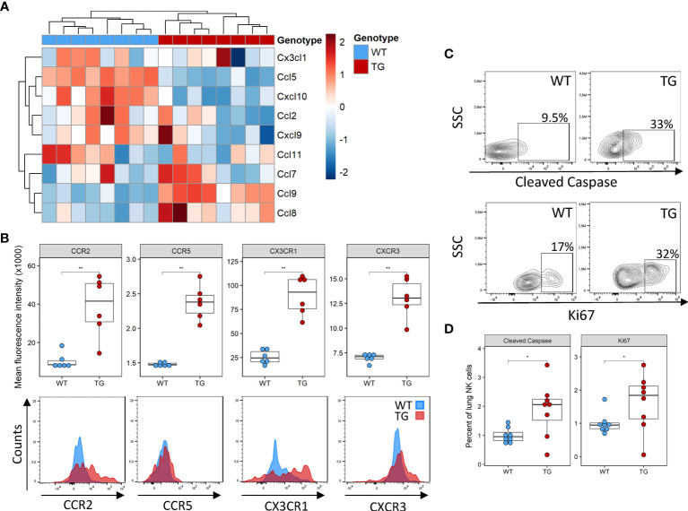
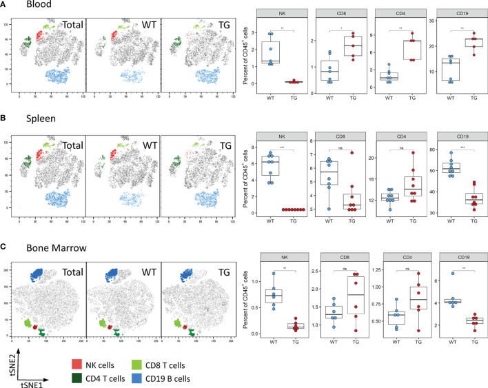
Natural killer (NK) cells play an important role in recognizing and killing pathogen-infected or malignant cells. Changes in their numbers or activation can contribute to several diseases and pathologies including systemic sclerosis (SSc), an autoimmune disease characterized by inflammation and tissue remodeling. In these patients, increased expression of the AP-1 transcription factor, Fra-2 was reported. In mice ectopic overexpression of Fra-2 (TG) leads to SSc with strong pulmonary fibrosis, pulmonary hypertension, and inflammation. Analysis of the underlying immune cell profile in the lungs of young TG mice, which do not yet show any signs of lung disease, revealed increased numbers of eosinophils and T cells but strongly reduced NK numbers. Therefore, we aimed to identify the cause of the absence of NK cells in the lungs of these mice and to determine the potential role of Fra-2 in NK development. Examination of inflammatory cell distribution in TG mice revealed similar NK deficiencies in the spleen, blood, and bone marrow. Deeper analysis of the WT and TG bone marrow revealed a potential NK cell developmental defect beginning at the preNKP stage. To determine whether this defect was cell-intrinsic or extrinsic, mixed bone marrow chimera and in vitro differentiation experiments were performed. Both experiments showed that the defect caused by Fra-2 was primarily cell-intrinsic and minimally dependent on the environment. Closer examination of surface markers and transcription factors required for NK development, revealed the expected receptor distribution but changes in transcription factor expression. We found a significant reduction in Nfil3, which is essential for the transition of common lymphoid cells to NK committed precursor cells and an AP-1 binding site in the promotor of this gene. In Summary, our data demonstrates that regulation of Fra-2 is essential for NK development and maturation, and suggests that the early NK dysfunction plays an important role in the pathogenesis of systemic sclerosis.
Keywords: AP-1; Fra-2; Natural killer (NK) cell; activator protein 1; differentiation; innate immunity.
Copyright © 2022 Schnoegl, Hochgerner, Gotthardt and Marsh.
Conflict of interest statement
The authors declare that the research was conducted in the absence of any commercial or financial relationships that could be construed as a potential conflict of interest.
Figures


Similar articles
-
The transcription factor Fra-2 regulates the production of extracellular matrix in systemic sclerosis.Arthritis Rheum. 2010 Jan;62(1):280-90. doi: 10.1002/art.25056. Arthritis Rheum. 2010. PMID: 20039427
-
Fra-2 Expression in Osteoblasts Regulates Systemic Inflammation and Lung Injury through Osteopontin.Mol Cell Biol. 2018 Oct 29;38(22):e00022-18. doi: 10.1128/MCB.00022-18. Print 2018 Nov 15. Mol Cell Biol. 2018. PMID: 30181393 Free PMC article.
-
Development of pulmonary fibrosis through a pathway involving the transcription factor Fra-2/AP-1.Proc Natl Acad Sci U S A. 2008 Jul 29;105(30):10525-30. doi: 10.1073/pnas.0801414105. Epub 2008 Jul 18. Proc Natl Acad Sci U S A. 2008. PMID: 18641127 Free PMC article.
-
Transcription factor Fra-2 and its emerging role in matrix deposition, proliferation and inflammation in chronic lung diseases.Cell Signal. 2019 Dec;64:109408. doi: 10.1016/j.cellsig.2019.109408. Epub 2019 Aug 29. Cell Signal. 2019. PMID: 31473307 Review.
-
Progress in the Study of Fra-2 in Respiratory Diseases.Int J Mol Sci. 2024 Jun 28;25(13):7143. doi: 10.3390/ijms25137143. Int J Mol Sci. 2024. PMID: 39000247 Free PMC article. Review.
Cited by
-
Single-cell RNA sequencing reveals altered immune and stromal landscape in primary and liver metastasis of gastric cancer.Biochem Biophys Rep. 2025 Jul 18;43:102163. doi: 10.1016/j.bbrep.2025.102163. eCollection 2025 Sep. Biochem Biophys Rep. 2025. PMID: 40727437 Free PMC article.
References
-
- Kee BL. Development of Natural Killer Cells and ILC1. Encyclopedia of Immunobiology Academic Press; (2016) 140–8. doi: 10.1016/B978-0-12-374279-7.04002-9 - DOI
Publication types
MeSH terms
Substances
LinkOut - more resources
Full Text Sources
Medical
Molecular Biology Databases
Research Materials
Miscellaneous