

Tactile and Ultrasound Image Fusion for Functional Assessment of the Female Pelvic Floor
- PMID: 35812797
- PMCID: PMC9262332
- DOI: 10.4236/ojog.2021.116063
Tactile and Ultrasound Image Fusion for Functional Assessment of the Female Pelvic Floor
Abstract
Introduction: The true etiology of pelvic organ prolapse and urinary incontinence and variations observed among individuals are not entirely understood. Tactile (stress) and ultrasound (anatomy, strain) image fusion may furnish new insights into the female pelvic floor conditions. This study aimed to explore imaging performance and clinical value of vaginal tactile and ultrasound image fusion for characterization of the female pelvic floor.
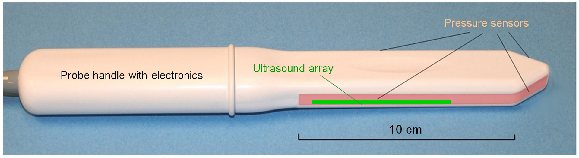
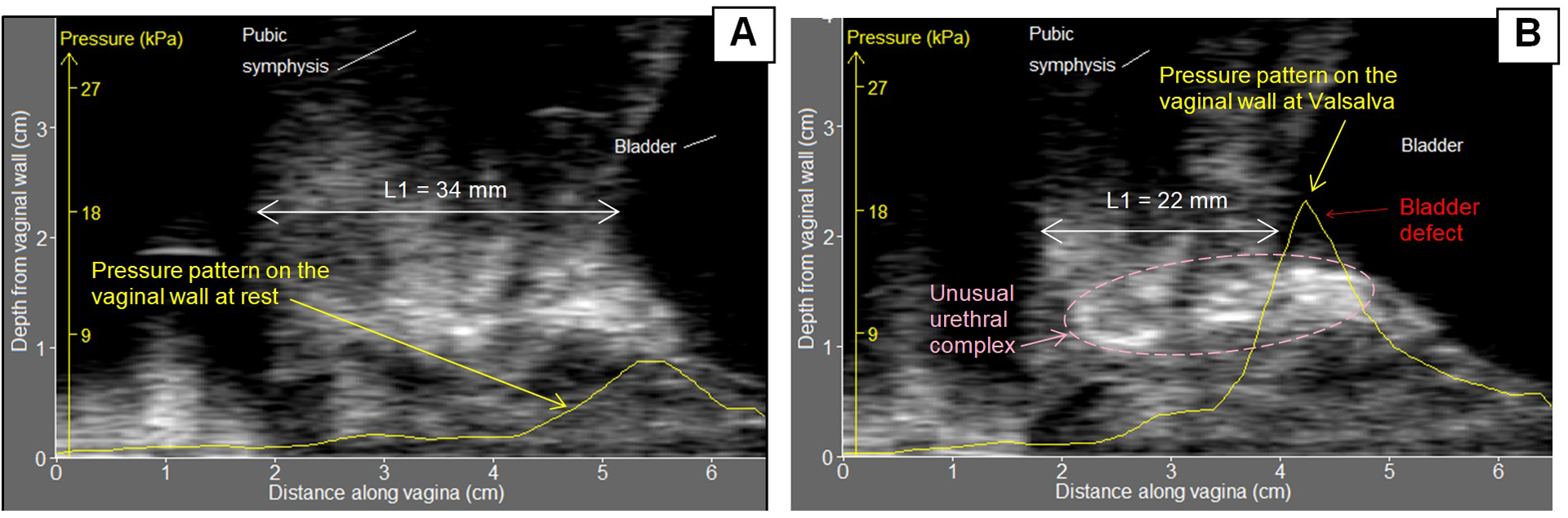
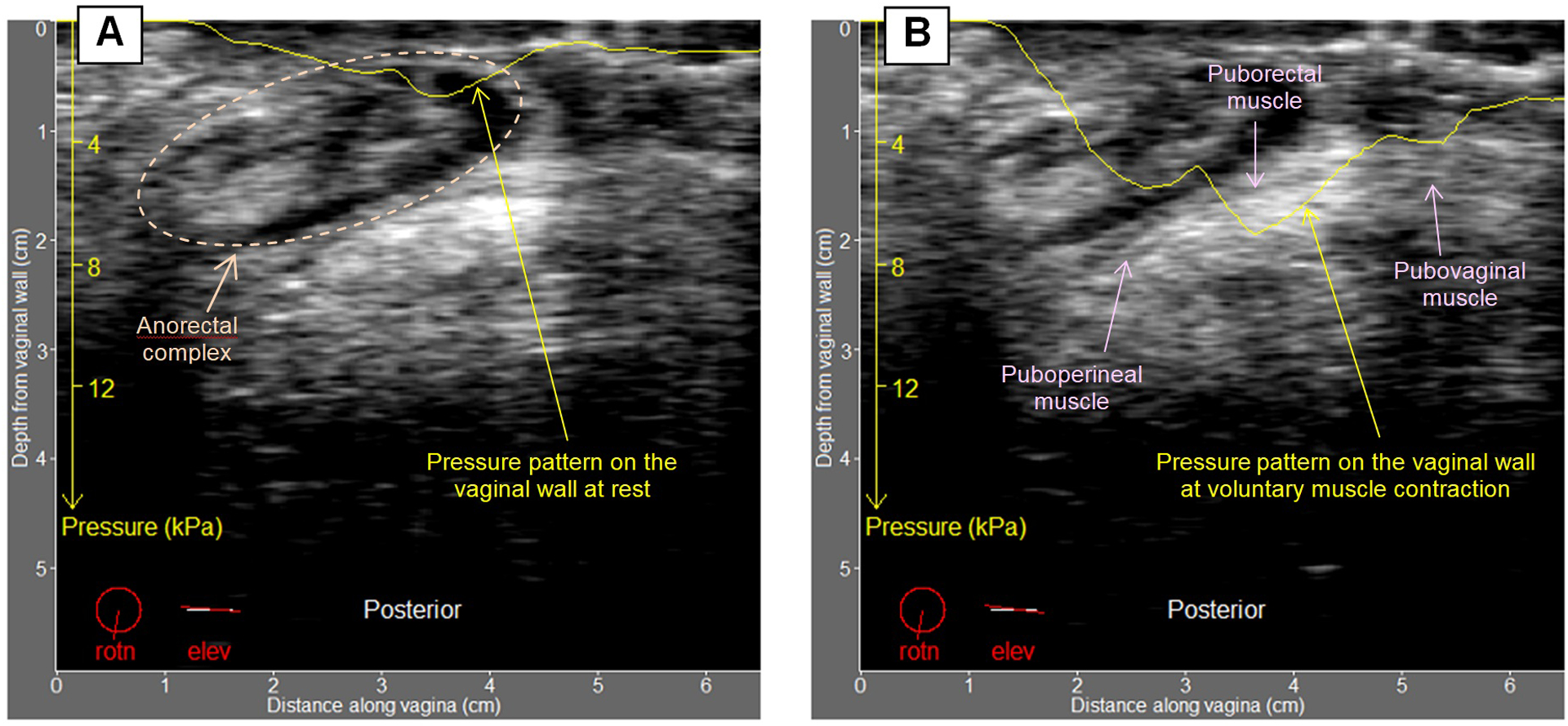
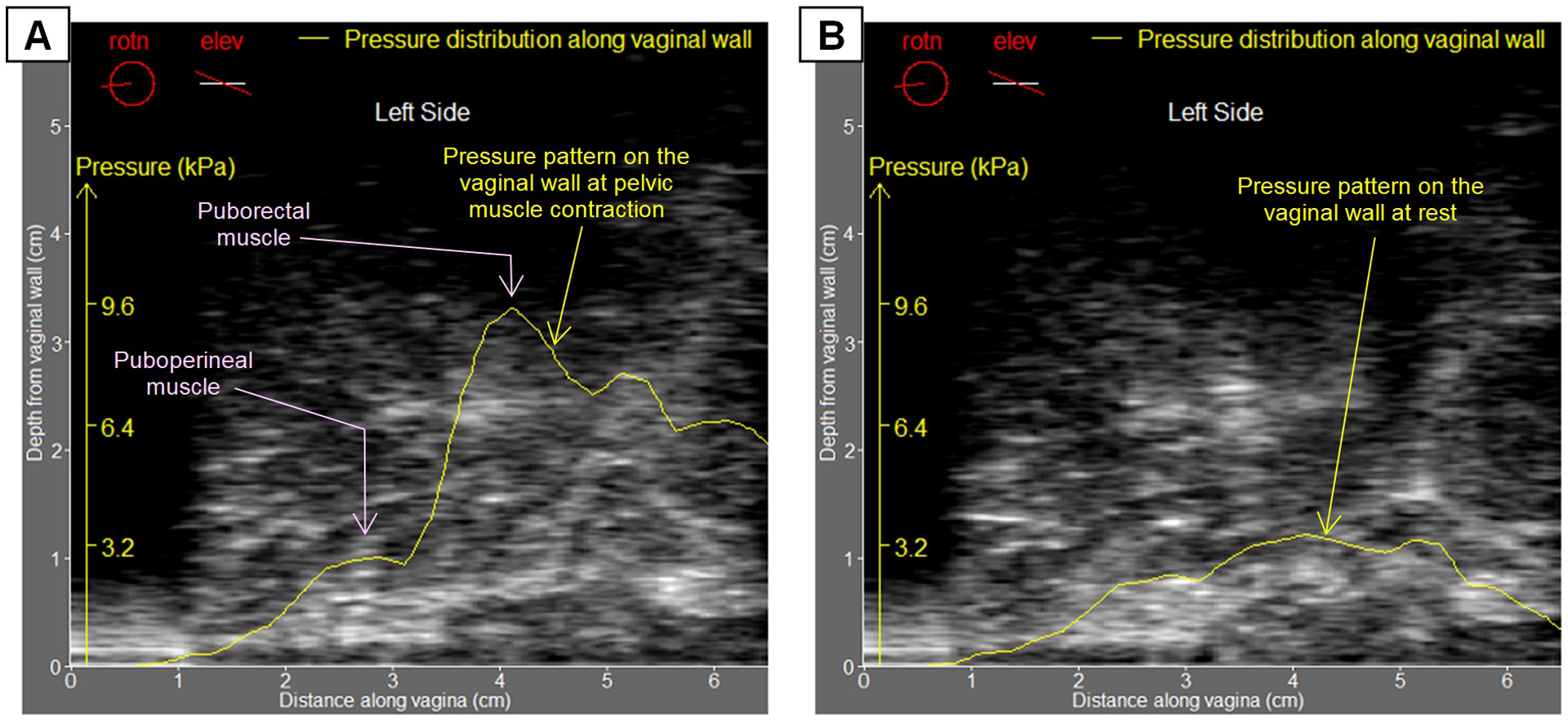
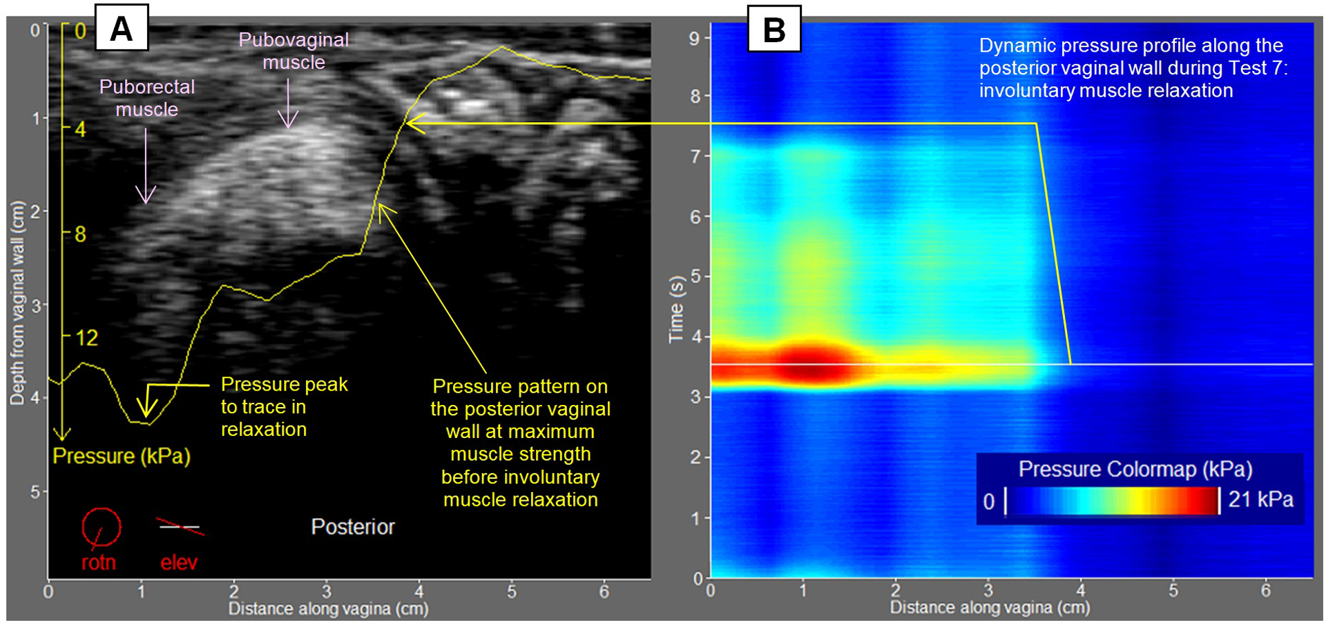
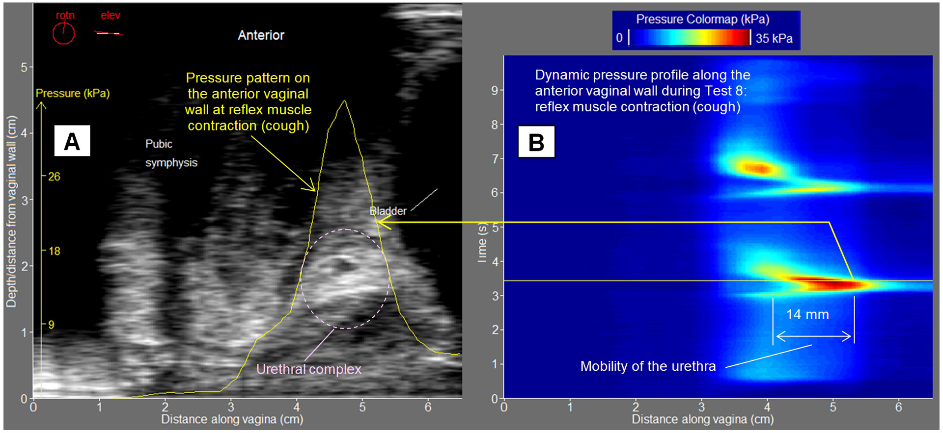
Methods: A novel probe with 96 tactile and 192 ultrasound transducers was designed. Women scheduled for a urogynecological visit were considered eligible for enrollment to observational study. Intravaginal tactile and ultrasound images were acquired for vaginal wall deformations at probe insertion, elevation, rotation, Valsalva maneuver, voluntary contractions, involuntary relaxation, and reflex pelvic muscle contractions. Biomechanical mapping has included tactile/ultrasound imaging and functional imaging.
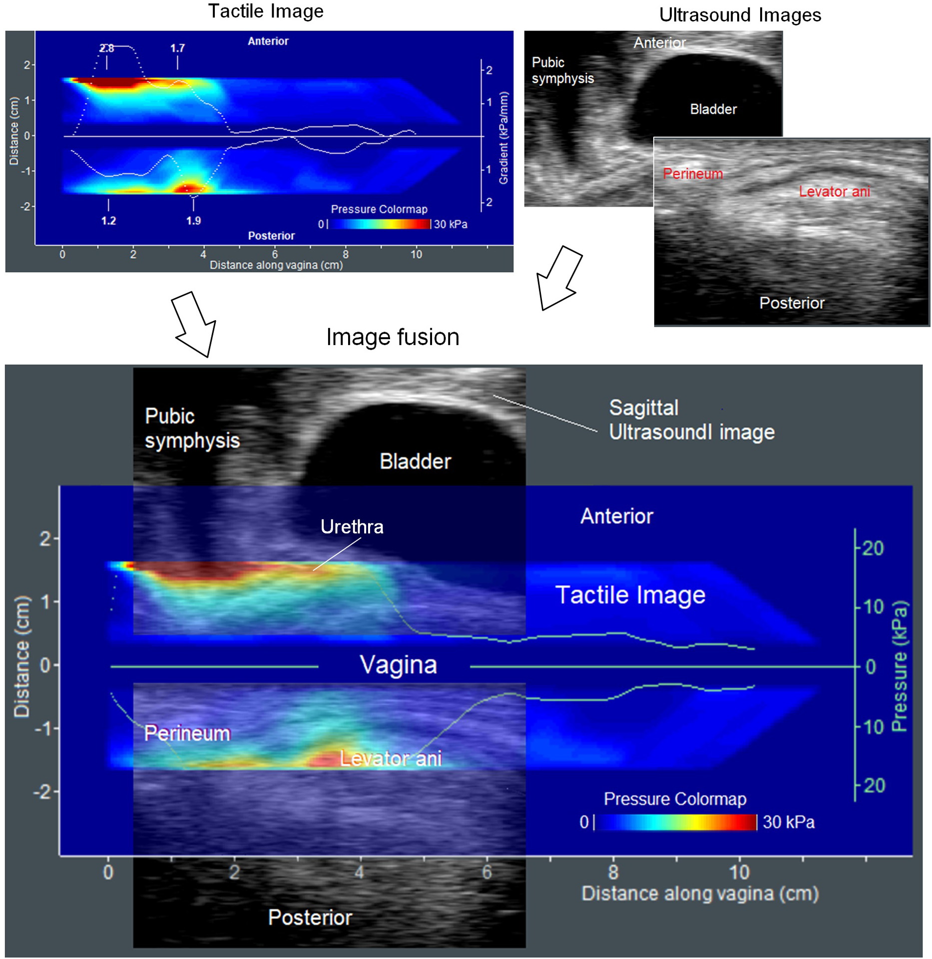
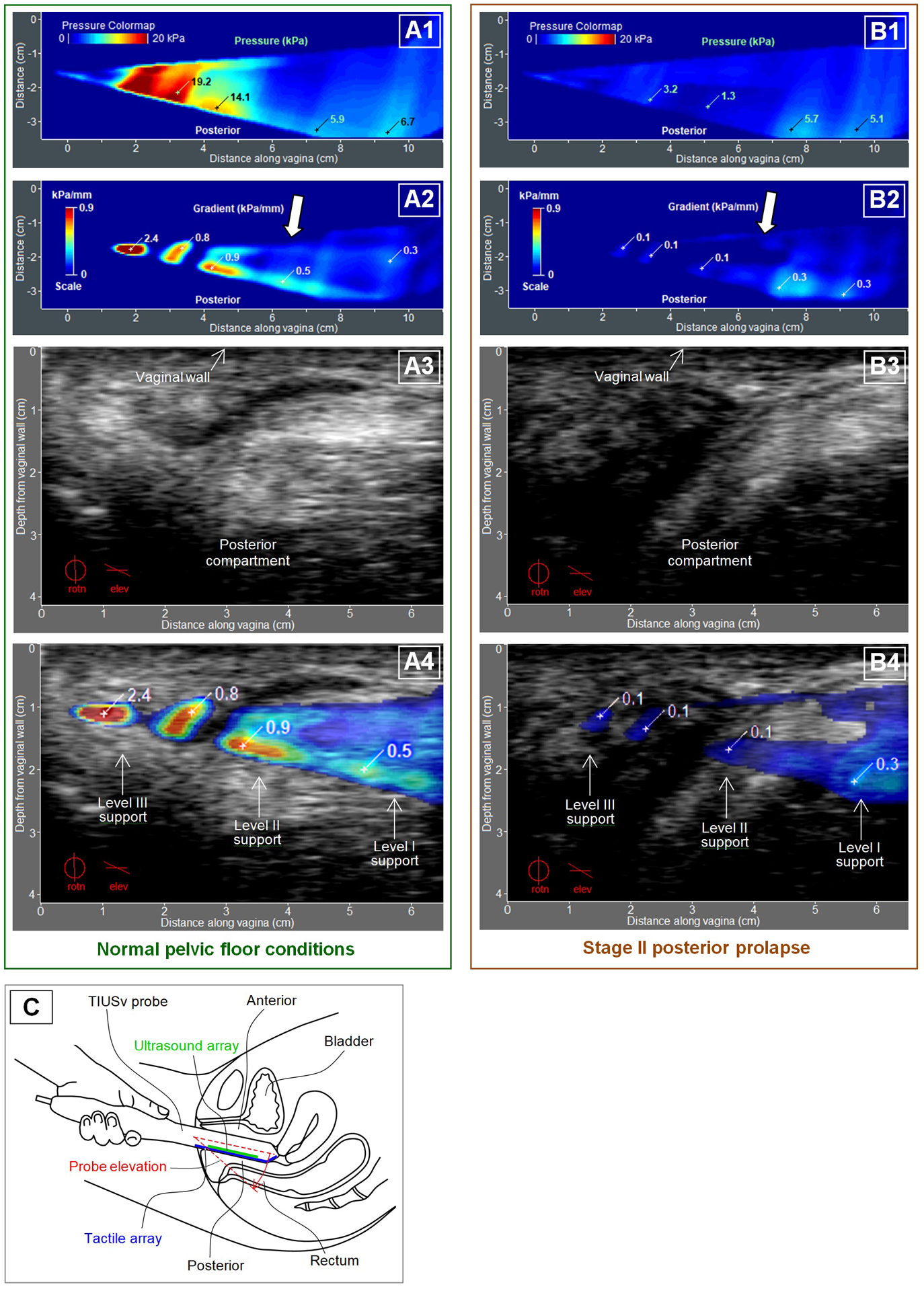
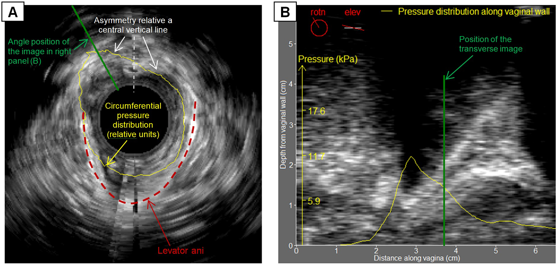
Results: Twenty women were successfully studied with the probe. Tactile and ultrasound images for tissues deformation as well as functional images were recorded. Tactile (stress) and ultrasound (strain) images allowed creation of stress-strain maps for the tissues of interest in absolute scale. Functional images allowed identification of active pelvic structures and their biomechanical characterization (anatomical measurements, contractive mobility and strength). Fusion of the modalities has allowed recognition and characterization of levator ani muscles (pubococcygeal, puborectal, iliococcygeal), perineum, urethral and anorectal complexes critical in prolapse and/or incontinence development.
Conclusions: Vaginal tactile and ultrasound image fusion provides unique data for biomechanical characterization of the female pelvic floor. Bringing novel biomechanical characterization for critical soft tissues/structures may provide extended scientific knowledge and improve clinical practice.
Keywords: Biomechanical Mapping; Pelvic Function; Pelvic Support; Tactile; Tissue Elasticity; Ultrasound.
Conflict of interest statement
Disclosure Egorov is a CEO and a minor shareholder of Advanced Tactile Imaging, Inc. Egorov has submitted a patent application related to the reported approach. Raalte is a minor shareholder of Advanced Tactile Imaging, Inc. Shobeiri reports no conflict of interest
Figures
References
-
- Iglesia CB, Smithling KR. Pelvic Organ Prolapse. Am Fam Physician. 2017; 96(3): 179–85. - PubMed
-
- Al-Mukhtar Othman J, Åkervall S, Milsom I, Gyhagen M. Urinary incontinence in nulliparous women aged 25–64 years: a national survey. Am J Obstet Gynecol. 2017; 216(2): 149.e1–149.e11. - PubMed
-
- Shobeiri S 2D/3D Endovaginal and Endoanal Instrumentation and Techniques. In: Practical Pelvic Floor Ultrasonography: A Multi-compartmental Approach to 2D/3D/4D Ultrasonography of Pelvic Floor. New York: Springer-Verlag; 2017. p. 19–44.
Grants and funding
LinkOut - more resources
Full Text Sources