

Morning glory disc anomaly associated with moyamoya disease and pituitary stalk duplication
- PMID: 35813587
- PMCID: PMC9259472
- DOI: 10.1016/j.ajoc.2022.101632
Morning glory disc anomaly associated with moyamoya disease and pituitary stalk duplication
Abstract
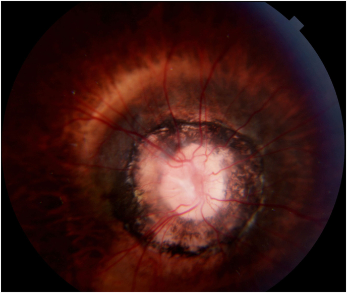
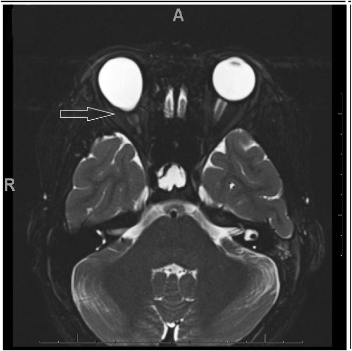
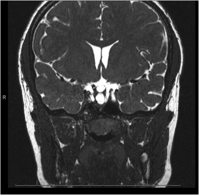
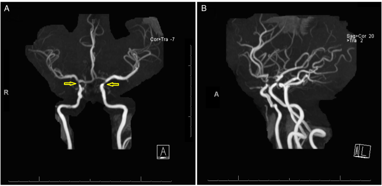
Purpose: We report a case of a 10-year-old with Moring glory disc anomaly (MGDA) associated with Moyamoya disease and pituitary stalk duplication.
Observations: A 10-year-old Asian child presented with decreased vision in the right eye and bilateral nystagmus. Both dilated fundus exam and magnetic resonance imaging (MRI) of the orbit confirmed MGDA of the right eye. MRI of the brain demonstrated duplication of the pituitary stalk. Magnetic resonance angiography (MRA) of the brain revealed bilateral severe narrowing (greater on the right side) of the distal supraclinoid internal carotid arteries with bilateral reconstitution at the carotid terminus and prominent collaterals, suggestive of Moyamoya disease.
Conclusions: Patients with MGDA should undergo neuroimaging due to the associated central nervous system (CNS) anomalies.
Keywords: Morning glory disc anomaly; Moyamoya disease.
© 2022 Published by Elsevier Inc.
Conflict of interest statement
The authors have no relevant conflicts of interest to disclose with this manuscript.
Figures
Similar articles
-
Pituitary stalk duplication in association with moya moya disease and bilateral morning glory disc anomaly - broadening the clinical spectrum of midline defects.J Neurol. 2008 Jun;255(6):885-90. doi: 10.1007/s00415-008-0799-5. Epub 2008 Mar 20. J Neurol. 2008. PMID: 18350354
-
Internal carotid artery origin of the anterior cerebral artery: A rare anatomic intracranial arterial variation in a child with morning glory disc anomaly and moyamoya vascular pattern; case report and review of literature.Brain Circ. 2020 Jun 26;6(2):133-138. doi: 10.4103/bc.bc_10_20. eCollection 2020 Apr-Jun. Brain Circ. 2020. PMID: 33033785 Free PMC article.
-
Morning glory syndrome with Moyamoya disease: A rare association with role of imaging.Indian J Radiol Imaging. 2018 Apr-Jun;28(2):165-168. doi: 10.4103/ijri.IJRI_219_17. Indian J Radiol Imaging. 2018. PMID: 30050238 Free PMC article.
-
Postoperative follow-up of a case of atypical morning glory syndrome associated with persistent fetal vasculature.BMC Ophthalmol. 2019 Jul 16;19(1):150. doi: 10.1186/s12886-019-1154-6. BMC Ophthalmol. 2019. PMID: 31311513 Free PMC article. Review.
-
Moyamoya Disease Associated With Morning Glory Disc Anomaly and Other Ophthalmic Findings: A Mini-Review.Front Neurol. 2020 May 15;11:338. doi: 10.3389/fneur.2020.00338. eCollection 2020. Front Neurol. 2020. PMID: 32499749 Free PMC article. Review.
Cited by
-
Pituitary Stalk Duplication: A Radiological Surprise in a Child With Short Stature.AACE Clin Case Rep. 2023 Jun 30;9(5):166-169. doi: 10.1016/j.aace.2023.06.004. eCollection 2023 Sep-Oct. AACE Clin Case Rep. 2023. PMID: 37736324 Free PMC article.
-
Vitrectomy Combined With Gas Tamponade for the Treatment of Morning Glory Syndrome With Rhegmatogenous Retinal Detachment: A Case Report.Cureus. 2025 Feb 5;17(2):e78550. doi: 10.7759/cureus.78550. eCollection 2025 Feb. Cureus. 2025. PMID: 40062134 Free PMC article.
-
(What's the story) morning glory? MRI findings in morning glory disc anomaly.Neuroradiology. 2024 Jul;66(7):1225-1233. doi: 10.1007/s00234-024-03375-2. Epub 2024 May 8. Neuroradiology. 2024. PMID: 38717474
-
Progress in investigating pituitary stalk lesions: A review.Medicine (Baltimore). 2025 Jan 10;104(2):e41232. doi: 10.1097/MD.0000000000041232. Medicine (Baltimore). 2025. PMID: 39792770 Free PMC article. Review.
References
-
- Quah B.L., Hamilton J., Blaser S., Héon E., Tehrani N.N. Morning glory disc anomaly, midline cranial defects and abnormal carotid circulation: an association worth looking for. Pediatr Radiol. 2005;35(5):525–528. - PubMed
-
- Harasymowycz P., Chevrette L., Décarie J.C., et al. Morning glory syndrome: clinical, computerized tomographic, and ultrasonographic findings. J Pediatr Ophthalmol Strabismus. 2005;42(5):290–295. - PubMed
-
- Ghaffari-Rafi A., Ghaffari-Rafi S., Leon-Rojas J. Socioeconomic and demographic disparities of moyamoya disease in the United States. Clin Neurol Neurosurg. 2020;192(105719) - PubMed
Publication types
LinkOut - more resources
Full Text Sources
