. 2022 Jun;14(6):1784-1793.
doi: 10.21037/jtd-21-1485.

A novel angiographic classification of pseudoaneurysms of the pulmonary chronic inflammatory cavity based on selective angiograms and therapeutic implications

Affiliations

A novel angiographic classification of pseudoaneurysms of the pulmonary chronic inflammatory cavity based on selective angiograms and therapeutic implications

Shengli Chen et al. J Thorac Dis. 2022 Jun.

Abstract

Background: Hemoptysis is a common clinical symptom. In the chronic tuberculosis cavity and chronic necrotizing pneumonia cavity, pseudoaneurysms (Pas) easily form and are prone to massive hemoptysis and repeated hemoptysis and can even endanger patient's life. However, it remains to be further analyzed whether Pas of the pulmonary chronic inflammatory cavity selectively affect the peripheral pulmonary branches. This study is based on selective angiography to classify peripheral pulmonary arterial Pas (PAPs) of the pulmonary chronic inflammatory cavity and to determine treatment options for PAPs, thereby guiding individualized clinical treatment.

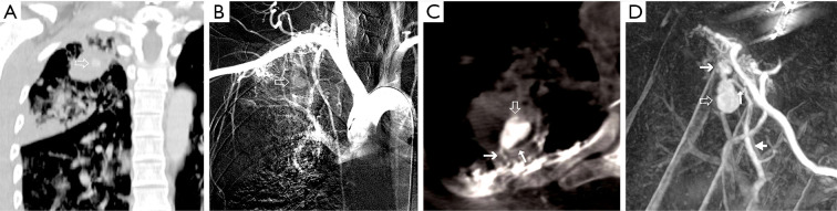
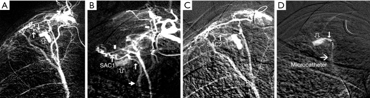
Methods: Angiographic data of 392 noncancer patients undergoing hemoptysis were retrospectively analyzed. All of the patients underwent pulmonary and selective pulmonary angiography and bronchial and nonbronchial systemic collateral arterial angiography. A total of 9 patients had Pas of the pulmonary chronic inflammatory cavity, and a pseudoaneurysm systemic artery collateral (Pasac), inflow and outflow sections of the parent vessels, and direction of blood flow in the parent vessels were clearly observed with digital subtraction angiography (DSA) and/or C-arm cone-beam flat-panel detector computed tomography angiography (CBCTA). Patients with underlying disease had pulmonary tuberculosis (n=8) or lung abscess (n=1). The angiographic types of Pas were analyzed.

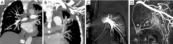
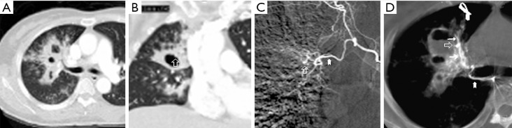
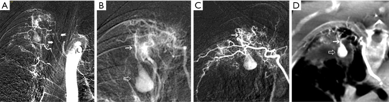
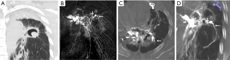
Results: Eight patients with chronic pulmonary tuberculosis and 1 patient with a necrotizing pneumonia cavity in the convalescent period were included in the study. Pas of the pulmonary chronic inflammatory cavity presented the following types: (I) pulmonary artery pseudoaneurysm (PAPa) (n=2 cases); (II) body arterial Pa (n=3 cases); and (III) systemic-pulmonary anastomosis Pa. Each type could be divided into two subtypes (n=4 cases). In nine cases, embolization and hemostasis were technically and clinically successful.

Conclusions: Pas of the pulmonary chronic inflammatory cavity are diverse (especially in cases of pulmonary tuberculosis). Angiographic typing plays a guiding role in the selection of an embolization strategy.

Keywords: Pseudoaneurysm (Pa); angiography; cavity; embolization; hemoptysis.


Conflict of interest statement

Conflicts of Interest: All authors have completed the ICMJE uniform disclosure form (available at https://jtd.amegroups.com/article/view/10.21037/jtd-21-1485/coif). The authors have no conflicts of interest to declare.

Figures

Figure 1
Figure 1
Chronic cavity Pa of the lung abscess on the left upper lung tip posterior segment (case 2). Chest enhanced computed tomographic PA phase (A) and left pulmonary angiography (C), Pa not revealed. Chest enhanced computed tomographic aortic stage (B) and left subclavian angiography (D), the Pa and its parent vessel of anterior subsegmental PAbs of the posterior left upper pulmonary apex were retrogradely revealed centripetal filling retrogradely. Super selective angiography confirmed that three branches of the left subclavian artery (three block diagrams; D) feeding the Pa, from their hyperplastic vascular network emitted two collateral branches and anastomoses with the two distal branches of the parent vessel of the Pa. The arrows in (B) are defined as the retrogradely revealed Pa and its parent vessel of anterior subsegmental PAbs of the posterior left upper pulmonary apex during enhanced computed tomographic aortic stage; the arrows in (D) are defined as the retrogradely revealed Pa and its parent vessel of anterior subsegmental PAbs of the posterior left upper pulmonary apex during super selective left subclavian angiography. Pa, pseudoaneurysm; PA, pulmonary artery; PAb, pulmonary artery branch.
Figure 2
Figure 2
Tuberculosis cavity Pa in the anterior segment of the right upper lobe (case 3). Chest enhancement computed tomographic (A) showed multiple tuberculous cavities in the upper right pulmonary, one of which had a significantly enhanced small nodule in the upper-medial wall (B). The right bronchial intercostal artery trunk angiography (C) and CBCTA axis MPR image (D), showed that the branches of the right BA formed a small oval Pa on the upper-medial wall of the cavity. The arrow in (B) is defined as a significantly enhanced small nodule in one of tuberculous cavities in the upper right pulmonary during enhancement computed tomographic; the arrows in (C) are defined as the right bronchial intercostal artery trunk angiography; the arrows in (D) are defined as the right bronchial intercostal artery trunk CBCTA axis MPR image. Pa, pseudoaneurysm; CBCTA, C-arm cone-beam flat-panel detector computed tomography angiography; MPR, multiplanar reconstruction; BA, bronchial artery.
Figure 3
Figure 3
The Pa of the right upper pulmonary posterior segment of the chronic tuberculous cavity (case 4). PA angiography showed decreased branches of the right upper PA (no show). Thoracic aortic angiography revealed a water droplet shape Pa in the right upper pulmonary (A). The right third and fourth intercostal arteriography (B,C) and fourth intercostal artery CBCTA MPR image (D), revealed a systemic artery collateral vessel enter the cavity wall, and the terminal damage forms a Pa, the collateral vessel come from hyperplasia vascular network of the right 3rd and 4th intercostal arteries. Systemic-PA anastomosis occurred in fibrous plaque that adjacent cavity (A,B). The arrows in (A) are defined as a water droplet shape Pa in the right upper pulmonary during thoracic aortic angiography; the arrows in (B) are defined as the right third intercostal arteriography; the arrows in (C) are defined as the right fourth intercostal arteriography; the arrows in (D) are defined as the fourth intercostal artery CBCTA MPR image. Pa, pseudoaneurysm; PA, pulmonary artery; CBCTA, C-arm cone-beam flat-panel detector computed tomography angiography; MPR, multiplanar reconstruction.
Figure 4
Figure 4
Tuberculosis cavity Pa in the superior segment of left lower lobe (case 7). Thoracic no-enhanced computed tomographic scan showed apico-posterior segment of the left upper lobe damaged and atelectasis, and there were a tuberculous cavity in the dorsal segment of the compensatory dilated left lower lobe, and a PAb connects the cavity the CMN (A). Selective angiography showed that a total of two left bronchial arteries and a left lower pulmonary ligament artery were the blood supplying vessels of Pa in the CMN, and the branches of the three arteries formed a hyperplasia vessels network and sent out a collateral vessel to form the inflow vessel of the Pa. One of the left bronchial angiography (B) and the CBCTA appropriate angle MPR images (C,D), revealed that this inflow vessel of the Pa walk windingly along medial-anterior-inferior wall of the cavity, and anastomosis with a small branch of PA to form the parent vessel of Pa, the parent vessel herniated into the CMN at the anastomosis and formed the Pa. The outflow PAb of the Pa showed retrograde centripetal filling. The arrows in (B) are defined as the left bronchial selective angiography; the arrows in (C) are defined as the left bronchial CBCTA images; the arrows in (D) are defined as the left bronchial MPR images. Pa, pseudoaneurysm; PAb, pulmonary artery branch; CMN, cavity mural nodule; CBCTA, C-arm cone-beam flat-panel detector computed tomography angiography; MPR, multiplanar reconstruction; PA, pulmonary artery.
Figure 5
Figure 5
Tuberculosis cavity Pa in the posterior superior lung (case 8). Enhanced computed tomographic reveal a high density node in the left upper lung effusion tuberculosis cavity (A). The posterior segment PA of the right upper lobe no fill during right pulmonary angiography (no shown). Thoracic aortography revealed the Pa in cavity as a gourd shape (B). Right second and third intercostal arterial trunk angiography (no show), and adaptive angle MPR (C) and MIP (D) reconstruction images of CBCTA, revealed that the branches of the right 2nd and 3rd intercostal arteries formed hyperplasia vascular network, and emitting collateral vessel to form the inflow vessel of the Pa. It anastomosis with the PAb on the cavity wall, and herniated into cavity to form a Pa at anastomosis site. The outflow vessel PAbs retrograde centripetal filled. The arrow in (A) is defined as a high density node in the left upper lung effusion tuberculosis cavity during enhanced computed tomographic; the arrow in (B) is defined as a gourd shape Pa in cavity during thoracic aortography; the arrows in (C) are defined as the branches of the right 2nd and 3rd intercostal arteries formed hyperplasia vascular network revealed by adaptive angle MPR; the arrows in (D) are defined as the branches of the right 2nd and 3rd intercostal arteries formed hyperplasia vascular network revealed by MIP. Pa, pseudoaneurysm; PA, pulmonary artery; MPR, multiplanar reconstruction; MIP, maximum density projection reconstruction; CBCTA, C-arm cone-beam flat-panel detector computed tomography angiography; PAb, pulmonary artery branch.
Figure 6
Figure 6
Tuberculosis cavity Pa in the right upper lung (case 9). Chest enhanced computed tomographic revealed irregular enhanced nodule in tuberculosis cavity of right upper pulmonary apex (no shown). Right costal-carotid trunk angiography (A) and CBCTA (no shown), and its branch right upper intercostal artery (B) and right second intercostal artery (C) angiography, revealed their collateral branches SAC1 and SAC2 preferentially supplied blood to the transverse and longitudinal parts of the “7”-shaped Pa, respectively. The inflowing blood vessel SAC1 of Pa presents as a loose spring, and anastomoses on the wall of the cavity with a flexible branch of the outflow PA, to form the parent vessel of Pa, this outflow PAb manifested as retrograde centripetal filling. The inflow vessel SAC2 filled the longitudinal portion of the Pa, and then slowly flowed out through the PAb that anastomosed with SAC1. The feeding systemic arteries of Pa were embolized using PVA particles, and the outflowing PA vessel was super selective embolized using micro coil near the Pa neck through right femoral vein catheterization (D). The arrows in (A) are defined as the right costal-carotid trunk angiography; the arrows in (B) are defined as the right upper intercostal artery angiography; the arrows in (C) are defined as the right second intercostal artery angiography; the arrows in (D) are defined as the outflowing PA vessel was super selective embolized using micro coil near the Pa neck through right femoral vein catheterization. SAC1, systemic artery collateral 1; SAC2, systemic artery collateral 2; Pa, pseudoaneurysm; CBCTA, C-arm cone-beam flat-panel detector computed tomography angiography; PA, pulmonary artery; PAb, pulmonary artery branch; PVA, polyvinyl alcohol.

Similar articles

References

    1. Sbano H, Mitchell AW, Ind PW, et al. Peripheral pulmonary artery pseudoaneurysms and massive hemoptysis. AJR Am J Roentgenol 2005;184:1253-9. 10.2214/ajr.184.4.01841253 - DOI - PubMed
    1. Remy J, Lemaitre L, Lafitte JJ, et al. Massive hemoptysis of pulmonary arterial origin: diagnosis and treatment. AJR Am J Roentgenol 1984;143:963-9. 10.2214/ajr.143.5.963 - DOI - PubMed
    1. Tsukada J, Hasegawa I, Torikai H, et al. Interventional therapeutic strategy for hemoptysis originating from infectious pulmonary artery pseudoaneurysms. J Vasc Interv Radiol 2015;26:1046-51.e1. 10.1016/j.jvir.2015.04.002 - DOI - PubMed
    1. Shin S, Shin TB, Choi H, et al. Peripheral pulmonary arterial pseudoaneurysms: therapeutic implications of endovascular treatment and angiographic classifications. Radiology 2010;256:656-64. 10.1148/radiol.10091416 - DOI - PubMed
    1. Kierse R, Jensen U, Helmberger H, et al. Value of multislice CT in the diagnosis of pulmonary artery pseudoaneurysm from Swan-Ganz catheter placement. J Vasc Interv Radiol 2004;15:1133-7. 10.1097/01.RVI.0000133223.71673.22 - DOI - PubMed