Review
. 2022 Jun;14(6):2309-2325.
doi: 10.21037/jtd-22-182.

Systolic anterior motion of the mitral valve in hypertrophic cardiomyopathy: a narrative review

Affiliations
Review

Systolic anterior motion of the mitral valve in hypertrophic cardiomyopathy: a narrative review

Sarah A Guigui et al. J Thorac Dis. 2022 Jun.

Abstract

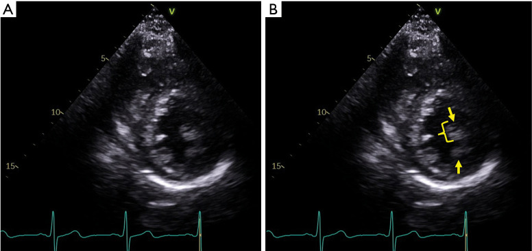
Background and objective: The prevalence of hypertrophic cardiomyopathy (HCM) is estimated to be 1 in 200 to 500 individuals, with systolic anterior motion (SAM) of the mitral valve (MV) and left ventricular outflow tract (LVOT) obstruction present in 60% to 70%. In this narrative review, we aim to elucidate the pathophysiology of SAM-septal contact and LVOT obstruction in HCM by presenting a detailed review on the anatomy of the MV apparatus in HCM, examining the various existing theories pertaining to the SAM phenomenon as supported by cardiac imaging, and providing a critical assessment of management strategies for SAM in HCM.

Methods: A literature review was performed using PubMed, EMBASE, Ovid, and the Cochrane Library, of all scientific articles published through December 2021. A focus was placed on descriptive studies, reports correlating echocardiographic findings with pathologic diagnosis, and outcomes studies.

Key content and findings: The pathophysiology of SAM involves the complex interplay between HCM morphology, MV apparatus anatomic abnormalities, and labile hemodynamic derangements. Echocardiography and cardiac magnetic resonance (CMR) vector flow mapping have identified drag forces, as opposed to the "Venturi effect", as the main hydraulic forces responsible for SAM. The degree of mitral regurgitation with SAM is variable, and its severity is correlated with degree of LVOT obstruction and outcomes. First line therapy for the amelioration of SAM and LVOT obstruction is medical therapy with beta-blockers, non-dihydropyridine calcium-channel blockers, and disopyramide, in conjunction with lifestyle modifications. In refractory cases septal reduction therapy is performed, which may be combined with a 'resect-plicate-release' procedure, anterior mitral leaflet extension, surgical edge-to-edge MV repair, anterior mitral leaflet retention plasty, or secondary chordal cutting.

Conclusions: Recent scientific advances in the field of HCM have allowed for a maturation of our understanding of the SAM phenomenon. Cardiac imaging plays a critical role in its diagnosis, treatment, and surveillance, and in our ability to apply the appropriate therapeutic regimens. The increasing prevalence of HCM places an emphasis on continued basic and clinical research to further improve outcomes for this challenging population.

Keywords: Hypertrophic cardiomyopathy (HCM); left ventricular outflow tract obstruction; mitral valve regurgitation; systolic anterior motion (SAM).


Conflict of interest statement

Conflicts of Interest: All authors have completed the ICMJE uniform disclosure form (available at https://jtd.amegroups.com/article/view/10.21037/jtd-22-182/coif). CGM serves as an unpaid editorial board member of Journal of Thoracic Disease from February 2021 to January 2023. The other authors have no conflicts of interest to declare.

Figures

Figure 1
Figure 1
Asymmetric interventricular septal thickening in hypertrophic cardiomyopathy. (A) A parasternal long-axis view at end-diastole; note the sigmoid septum morphology; (B) the solid line measures the maximum thickness of the basal interventricular septum at 20 mm, the double arrowhead highlights left ventricular outflow tract narrowing, the asterisk highlights a thickened mitral valve with an anteriorly-displaced coaptation point, and the single arrowhead points to short and fibrotic chordae tendinae.
Figure 2
Figure 2
Elongated mitral valve leaflets in hypertrophic cardiomyopathy. (A) A mid-diastolic parasternal long-axis view; (B) the double-headed arrow highlights an elongated anterior mitral leaflet measuring 28 mm, the double-headed dashed arrow highlights an elongated posterior mitral valve leaflet measuring 16 mm, and the asterisk highlights a markedly thickened interventricular septum measuring 30 mm with a reverse curve morphology.
Figure 3
Figure 3
Papillary muscle abnormalities in hypertrophic cardiomyopathy. (A) An end-diastolic parasternal short-axis view; (B) the arrows point to a bifid anterolateral papillary muscle, with the bracket highlighting a hypertrophied anterior head measuring 13 mm in thickness.
Figure 4
Figure 4
Depiction of systolic anterior motion of the mitral valve by vector flow mapping in hypertrophic cardiomyopathy. In a normal patient (top panel), there is progression from an early systolic isovolumic vortex to an organized ejection flow in the LVOT without evidence of interaction with the mitral valve (A-C). In a patient with obstructive hypertrophic cardiomyopathy and elongated mitral valve leaflets (middle panel), the isovolumic vortex initiates SAM of the mitral valve by pushing on the posterior surface of the mitral valve and positioning it in the LVOT (D,E). Alternatively, SAM is initiated in the early ejection phase (F), whereby flow is first deflected posteriorly by the bulging septum and then pushes on the posterior mitral valve leaflet surface, as it courses from a posterior to an anterior direction. In the bottom panel, the high angle of attack of flow on the mitral valve makes drag, and not lift, the predominant physical force leading to SAM. Reproduced with permission from Ro et al. (36). LV, left ventricle; LA, left atrium; Ao, aorta; HCM, hypertrophic cardiomyopathy; LVOT, left ventricular outflow tract; SAM, systolic anterior motion.
Figure 5
Figure 5
Color Doppler assessment at late systole of left ventricular outflow tract obstruction and mitral regurgitation in obstructive hypertrophic cardiomyopathy. (A) An apical 3-chamber view with color Doppler assessment; (B) the left arrow points to eccentric posteriorly-directed mitral regurgitation secondary to systolic anterior motion of the mitral valve; note the ‘Coanda’ effect of the regurgitant jet. The right arrow points to flow acceleration and turbulence in the left ventricular outflow tract as systolic obstruction occurs.
Figure 6
Figure 6
Severe SAM of the mitral valve. (A) A late-systolic parasternal long-axis view of severe mitral valve SAM in a patient with hypertrophic cardiomyopathy and discrete upper septal thickening (sigmoid septum morphology); (B) findings in panel B include marked basal septal systolic thickening (solid line), SAM with anterior mitral leaflet-septal contact (solid arrow), and systolic obliteration of the left ventricular outflow tract (dashed arrow). SAM, systolic anterior motion.

References

    1. Teare D. Asymmetrical hypertrophy of the heart in young adults. Br Heart J 1958;20:1-8. 10.1136/hrt.20.1.1 - DOI - PMC - PubMed
    1. Morrow AG, Braunwald E. Functional aortic stenosis; a malformation characterized by resistance to left ventricular outflow without anatomic obstruction. Circulation 1959;20:181-9. 10.1161/01.CIR.20.2.181 - DOI - PubMed
    1. Wigle ED, Heimbecker RO, Gunton RW. Idiopathic ventricular septal hypertrophy causing muscular subaortic stenosis. Circulation 1962;26:325-40. 10.1161/01.CIR.26.3.325 - DOI - PubMed
    1. Shah PM, Gramiak R, Kramer DH. Ultrasound localization of left ventricular outflow obstruction in hypertrophic obstructive cardiomyopathy. Circulation 1969;40:3-11. 10.1161/01.CIR.40.1.3 - DOI - PubMed
    1. Maron BJ, Peterson EE, Maron MS, et al. Prevalence of hypertrophic cardiomyopathy in an outpatient population referred for echocardiographic study. Am J Cardiol 1994;73:577-80. 10.1016/0002-9149(94)90337-9 - DOI - PubMed