

Subretinal drusenoid deposits: An update
- PMID: 35813798
- PMCID: PMC9262011
- DOI: 10.4103/tjo.tjo_18_22
Subretinal drusenoid deposits: An update
Abstract
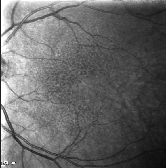
A wide spectrum of phenotypic manifestations characterizes age-related macular degeneration (AMD). Drusen is considered the hallmark of AMD and is located underneath the retinal pigment epithelium (RPE). In contrast, subretinal drusenoid deposits (SDDs), also known as reticular pseudodrusens, are located in the subretinal space, on top of the RPE. SDDs are poorly detected by clinical examination and color fundus photography. Multimodal imaging is required for their proper diagnosis. SDDs are topographically and functionally related to rods. SDDs cause a deep impairment in retinal sensitivity and dark adaptation. SDDs are dynamic structures that may grow, fuse with each other, or regress over time. An intermediate step in some eyes is the development of an acquired vitelliform lesion. The presence of SDD confers an eye a high risk for the development of late AMD. SDD leads to macular neovascularization, particularly type 3, geographic atrophy, and outer retinal atrophy.
Keywords: Age-related macular degeneration; outer retinal atrophy; pseudodrusen; reticular drusen; reticular macular disease; reticular pseudodrusen; retinal angiomatous proliferation; subretinal drusenoid deposit; type 3 macular neovascularization.
Copyright: © 2022 Taiwan J Ophthalmol.
Conflict of interest statement
The authors declare that there are no conflicts of interests of this paper.
Figures

References
-
- Steinberg JS, Göbel AP, Fleckenstein M, Holz FG, Schmitz-Valckenberg S. Reticular drusen in eyes with high-risk characteristics for progression to late-stage age-related macular degeneration. Br J Ophthalmol. 2015;99:1289–94. - PubMed
-
- Spaide RF, Ooto S, Curcio CA. Subretinal drusenoid deposits AKA pseudodrusen. Surv Ophthalmol. 2018;63:782–815. - PubMed
-
- Wightman AJ, Guymer RH. Reticular pseudodrusen: Current understanding. Clin Exp Optom. 2019;102:455–62. - PubMed
Publication types
LinkOut - more resources
Full Text Sources
Other Literature Sources