. 2022 Jun 23;4(4):fcac168.
doi: 10.1093/braincomms/fcac168. eCollection 2022.

Malin restoration as proof of concept for gene therapy for Lafora disease

Affiliations

Malin restoration as proof of concept for gene therapy for Lafora disease

Olga Varea et al. Brain Commun. .

Abstract

Lafora disease is a fatal neurodegenerative childhood dementia caused by loss-of-function mutations in either the laforin or malin gene. The hallmark of the disease is the accumulation of abnormal glycogen aggregates known as Lafora bodies (LBs) in the brain and other tissues. These aggregates are responsible for the pathological features of the disease. As a monogenic disorder, Lafora disease is a good candidate for gene therapy-based approaches. However, most patients are diagnosed after the appearance of the first symptoms and thus when LBs are already present in the brain. In this context, it was not clear whether the restoration of a normal copy of the defective gene (either laforin or malin) would prove effective. Here we evaluated the effect of restoring malin in a malin-deficient mouse model of Lafora disease as a proof of concept for gene replacement therapy. To this end, we generated a malin-deficient mouse in which malin expression can be induced at a certain time. Our results reveal that malin restoration at an advanced stage of the disease arrests the accumulation of LBs in brain and muscle, induces the degradation of laforin and glycogen synthase bound to the aggregates, and ameliorates neuroinflammation. These results identify malin restoration as the first therapeutic strategy to show effectiveness when applied at advanced stages of Lafora disease.

Keywords: Lafora disease; gene therapy; glycogen; neurodegeneration; neuroinflammation.


Figures

Graphical Abstract
Graphical Abstract
Figure 1
Figure 1
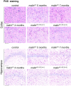
Malin restoration in young malinKO mice. PAS staining in brain. Representative images from control, 4-, 5-, and 7-month-old malinKO and malinKO+OE mice after 1 or 3 months of malin restoration. The upper panels show a cortical region and the lower panels the CA2/CA3 hippocampal region. Scale bar: 100 µm. n = 45 mice.
Figure 2
Figure 2
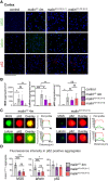
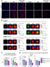
LB component analysis of young malinKO mice after malin restoration. (A) Representative immunofluorescence images from the cortical region obtained with anti-MGS (green), anti-laforin (green), or anti-p62 (red) stainings in combination with DAPI (blue). Control and 4-month-old malinKO (malinKO 4 m), malinKO+OE[4+1], and malinKO+OE[4+3] mice are shown. Scale bar: 100 µm. (B) Quantification of number of aggregates per area (mm2) detected by MGS, laforin, or p62 staining. ANOVA with Tukey’s multiple comparison test and unpaired t-test. MGS: control versus malinKO 4 months: P = 0.01, malinKO 4 months versus malinKO+OE[4+1]: P = 0.48, malinKO 4 months versus malinKO+OE[4+3]: P = 0.0092, malinKO+OE[4+1] versus malinKO+OE[4+3]: P = 0.21. Laforin: control versus malinKO 4 months: P = 0.0001, control versus malinKO+OE[4+1]: P = 0.021, malinKO 4 months versus malinKO+OE[4+1]: P = 0.022, malinKO 4 months versus malinKO+OE[4+3]: P = 0.009. p62: control versus malinKO 4 months: P = 0.01, malinKO 4 months versus malinKO+OE[4+1]: P = 0.23, malinKO 4 months versus malinKO+OE[4+3]: P = 0.69, malinKO+OE[4+1] versus malinKO+OE[4+3]: P = 0.51. P-value ≤ 0.05(*), P-value < 0.01 (**), and P-value < 0.001 (***), unpaired t-test. (C) Magnification of representative LBs found in 4-month-old malinKO mice (left) compared with malinKO+OE[4+3] mice (right). MGS and laforin (green) were co-stained with p62 (red), the overlay image for each one is shown (merge). Fluorescent profile along a line across the LB through its geometrical centre is shown in each case. The size of the fluorescent particle is indicated in each graphic. (D) Fluorescent intensity of MGS and laforin in p62-positive LBs from malinKO, malinKO+OE[4+1], and malinKO+OE[4+3] mice. ANOVA with Tukey’s multiple comparison test and unpaired t-test. MGS, malinKO 4 months versus malinKO+OE[4+1]: P ≤ 0.0001, malinKO 4 months versus malinKO+OE[4+3]: P ≤ 0.0001, malinKO+OE[4+1] versus malinKO+OE[4+3]: P = 0.9. Laforin: malinKO 4 months versus malinKO+OE[4+1]: P = 0.0006, malinKO 4 months versus malinKO+OE[4+3]: P = 0.013, malinKO+OE[4+1] versus malinKO+OE[4+3]: P = 0.77. p62: malinKO 4 months versus malinKO+OE[4+1]: P = 0.065, malinKO 4 months versus malinKO+OE[4+3]: P = 0.075, malinKO+OE[4+1] versus malinKO+OE[4+3]: P = 0.86. P-value ≤ 0.05(*), P-value < 0.01 (**), and P-value < 0.001 (***). Data are shown as mean ± SD. Each dot represents one mouse (n = 48 as indicated).
Figure 3
Figure 3
Malin restoration at an advanced stage of Lafora disease in malinKO mice. (A) PAS staining in brain. Representative images of cortical and CA2/CA3 hippocampal region from control, 11-month-old malinKO, 15-month-old malinKO, and malinKO+OE[11+4] mice are shown. ANOVA with Tukey’s multiple comparison test and unpaired t-test. Control versus malinKO 11 months: P = 0.00003, control versus malinKO 15 months: P = 0.0001, control versus malinKO+OE[11+4]: P = 0.0094, malinKO 11 months versus malinKO 15 months: P = 0.0032, malinKO 11 months versus malinKO+OE[11+4]: P = 0.96, malinKO 15 months versus malinKO+OE[11+4]: P = 0.0094. Scale bar: 100 µm. (B) Total brain glycogen was determined from the same groups (µg/mg of tissue). Each dot represents one mouse (n = 36).
Figure 4
Figure 4
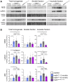
Soluble and insoluble brain fractions analysis. (A) Representative western blots of total brain homogenate, soluble, and insoluble fractions are shown for MGS, laforin, and p62. The images shown are cropped to show the band of interest, for full images see Supplementary material. Three samples from the same group were run for these images from a total of 3–6 mice per group. Loading control: LICOR—revert staining. (B) Quantifications of protein detection by western blot. Relative optical density units were related to loading control and normalized respect to control mice. Data are shown as mean ± SD. ANOVA with Tukey’s multiple comparison test and unpaired t-test. MGS total homogenate: control versus malinKO 11 months: P = 0.025, control versus malinKO 15 months: P = 0.02, malinKO 15 months versus malinKO[11+4]: P = 0.017, malinKO 11 months versus malinKO+OE[11+4]: P = 0.033, control versus malinKO+OE[11+4]: P = 0.028. MGS insoluble fraction: control versus malinKO 11 months: P = 0.01, control versus malinKO 15 months: P = 0.0001, malinKO 15 months versus malinKO+OE[11+4]: P = 0.015, malinKO 11 months versus malinKO+OE[11+4]: P = 0.026, control versus malinKO+OE[11+4]: P = 0.029. Laforin total homogenate: control versus malinKO 11 months: P = 0.0008, control versus malinKO 15 months: P = 0.02, malinKO 15months versus malinKO+OE[11+4]: P = 0.0076, malinKO 11 months versus malinKO+OE[11+4]: P = 0.08, control versus malinKO+OE[11+4]: P = 0.021. Laforin insoluble fraction: control versus malinKO 11 months: P = 0.04, control versus malinKO 15 months: P = 0.0094, malinKO 15 months versus malinKO+OE[11+4]: P = 0.0054, malinKO 11 months versus malinKO+OE[11+4]: P = 0.085, control versus malinKO+OE[11+4]: P = 0.0062. p62 total homogenate fraction: control versus malinKO 11 months: P = 0.105, control versus malinKO 15 months: P = 0.032, malinKO 11 months versus malinKO+OE[11+4]: P = 0.54, malinKO 15 months versus malinKO+OE[11+4]: P = 0.0511. p62 insoluble fraction: control versus malinKO 11 months: P = 0.04, control versus malinKO 15 months: P = 0.010, malinKO 11 months versus malinKO 15 months: P = 0.24, malinKO 11 months versus malinKO+OE[11+4]: P = 0.67, malinKO 15 months versus malinKO+OE[11+4]: P = 0.14. P-value ≤ 0.05(*), P-value < 0.01 (**), and P-value < 0.001 (***). Each dot represents one mouse (n = 36).
Figure 5
Figure 5
MGS, laforin, and p62 analysis in the hippocampus of malinKO mice after malin restoration at an advanced stage of Lafora disease. (A) Immunofluorescence images from the hippocampal region using anti-MGS (green), anti-laforin (red), or anti-p62 (red) antibodies combined with DAPI staining in each group. (B) Quantification of number of particles per area (mm2) in CA2/CA3 region stained with anti-MGS antibody. ANOVA with Tukey’s multiple comparison test. MGS total aggregates: control versus malinKO 11 months: P = 0.00038, control versus malinKO 15 months: P = 0.0008, control versus malinKO+OE[11+4]: P = 0.064, malinKO 11 months versus malinKO 15 months: P = 0.0697, malinKO 11 months versus malinKO+OE[11+4]: P = 0.0287, malinKO 15 months versus malinKO+OE[11+4]: P = 0.0006. nLBs: control versus malinKO 11 months: P = 0.0006, control versus malinKO 15 months: P = 0.0078, control versus malinKO+OE[11+4]: P = 0.1, malinKO 11 months versus malinKO 15 months: P = 0.0009, malinKO 11 months versus malinKO+OE[11+4]: P = 0.06, malinKO 15 months versus malinKO+OE[11+4]: P = 0.0175. CAL, control versus malinKO 11 months: P = 0.0023, control versus malinKO 15 months: P = 0.0008, control versus malinKO+OE[11+4]: P = 0.081, malinKO 11 months versus malinKO 15 months: P = 0.12, malinKO 11 months versus malinKO+OE[11+4]: P = 0.02, malinKO 15 months versus malinKO+OE[11+4]: P = 0.0011. (C) Quantification of nLBs and CALs from the hippocampus in each group. Quantification of number of particles per area (mm2) in CA2/CA3 hippocampal region stained with laforin (D) or p62 (E). ANOVA with Tukey’s multiple comparison test. Scale bar: 100 µm. In all graphics, data are shown as mean ± SD. P-value ≤ 0.05(*), P-value < 0.01 (**), and P-value < 0.001 (***). Each dot represents one mouse (n = 36 as indicated in the graphic).
Figure 6
Figure 6
Effect of late malin restoration on LB composition. (A) Representative immunofluorescent images from 15-month-old malinKO mice and malinKO+OE[11+4] mice. Co-stainings: MGS (green)/p62 (red) and laforin (green)/p62 (red), both combined with DAPI staining (blue), each channel is shown separately and as merge. Scale bar: 100 µm. (B) Two representative individual LBs and their immunofluorescent profile along a line crossing the geometrical centre are shown for each group. The profiles correspond to MGS (green)/p62 (red) or laforin (green)/p62 (red) combinations. Each channel and merge images are shown. The size of the p62-positive particle is indicated in each graphic. (C) Quantification of the percentage of double-positive MGS/p62 and laforin/p62 aggregates in each group. ANOVA with Tukey's multiple comparison test and unpaired t-test. MGS + p62: malinKO 11 months versus malinKO 15 months: P = 0.68, malinKO 11 months versus malinKO+OE[11+4]: P = 0.037, malinKO 15 months versus malinKO+OE[11+4]: 0.0146. Laforin + p62: malinKO 11 months versus malinKO 15 months: P = 0.002, malinKO 11 months versus malinKO+OE[11+4]: P ≤ 0.0001, malinKO 15 months versus malinKO+OE[11+4]: P = 0.02. (D) Fluorescent intensity of MGS and laforin in p62-positive aggregates. ANOVA with Tukey’s multiple comparison test and unpaired t-test. MGS mean Imax: malinKO 11 months versus malinKO 15 months: P = 0.76, malinKO 11 months versus malinKO+OE[11+4]: P = 0.0135, malinKO 15 months versus malinKO+OE[11+4]: 0.0273. Laforin mean Imax: malinKO 11 months versus malinKO 15 months: P = 0.1178, malinKO 11 months versus malinKO+OE[11+4]: P = 0.0004, malinKO 15 months versus malinKO+OE[11+4]: 0.0003. p62 mean Imax: malinKO 11 months versus malinKO 15 months: P = 0.37, malinKO 11 months versus malinKO+OE[11+4]: P = 0.311, malinKO 15 months versus malinKO+OE[11+4]: 0.67. P-value ≤ 0.05(*), P-value < 0.01 (**), and P-value < 0.001 (***). In all graphics, the data are shown as mean ± SD. Each dot represents one mouse.
Figure 7
Figure 7
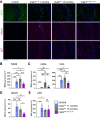
Inflammatory response after malin restoration at advanced stage of Lafora disease. (A) Astrogliosis and microgliosis were determined by immunostaining using anti-GFAP (red, combined with DAPI, blue) or anti-CD11b antibodies, respectively. Representative images from the hippocampus are shown. Scale bar: 500 µm. (B) Quantification of GFAP and CD11b stainings represented as percentage of positive pixel in the hippocampal region. N = 3 sections per mice, 36 mice. (C) RNA from total brain homogenates was analysed for quantification of the expression of pro-inflammatory genes by qPCR: IL-6, CXCL10. Graphics show the relative expression levels (2−ΔΔCt). Unpaired t-test. P-value ≤ 0.05(*), P-value < 0.01 (**), and P-value < 0.001 (***). CD11b: control versus malinKO[11+4] 11 months: P = 0.028, control versus malinKO 15 months: P = 0.0049, control versus malinKO+OE[11+4]: P = 0.0073, malinKO 11 months versus malinKO 15 months: P = 0.0037, malinKO 11 months versus malinKO+OE[11+4]: P = 0.18, malinKO 15 months versus malinKO+OE[11+4]: P = 0.064. GFAP: control versus malinKO 11 months: P = 0.002, control versus malinKO 15 months: P = 0.005, control versus malinKO+OE[11+4]: P = 0.035, malinKO 11 months versus malinKO 15 months: P = 0.75, malinKO 11 months versus malinKO+OE[11+4]: P = 0.82, malinKO 15 months versus malinKO+OE[11+4]: P = 0.86. IL6: control versus malinKO 11 months: P = 0.03, control versus malinKO 15 months: P = 0.004, control versus malinKO+OE[11+4]: P = 0.14, malinKO 11months versus malinKO 15 months: P = 0.018, malinKO 11 months versus malinKO+OE[11+4]: P = 0.8, malinKO 15 months versus malinKO+OE[11+4]: P = 0.018. CXCL10: control versus malinKO 11 months: P = 0.001, control versus malinKO 15 months: P = 0.0025, control versus malinKO+OE[11+4]: P = 0.1156, malinKO 11months versus malinKO 15 months: P = 0.0794, malinKO 11 months versus malinKO+OE[11+4]: P = 0.8, malinKO 15 months versus malinKO+OE[11+4]: P = 0.047. Each dot represents one mouse (n = 36 mice). Data are shown as mean ± SD.
Figure 8
Figure 8
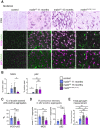
Effect of late malin expression on skeletal muscle. (A) PAS staining and immunofluorescent images from quadriceps muscles are shown for each group. Co-stainings of MGS/p62 were performed. For simplicity, MGS and p62 are shown independently. MGS (magenta, middle panels) and p62 (magenta, lower panels) were combined with agglutinin (WGA) to visualize the fibres (green). Scale bar: 100 µm. (B) The amount of MGS and p62 in the tissue was quantified as percentage of positive pixel in each group and represented as mean ± SD. Unpaired t-test, MGS % positive pixel: malinKO 15 months versus malinKO+OE[11+4]: P = 0.0022; p62% positive pixel: malinKO 15 months versus malinKO+OE[11+4]: P = 0.88. P-value ≤ 0.05(*), P-value < 0.01 (**), and P-value < 0.001 (***). Unpaired t-test, P-value ≤ 0.05(*), P-value < 0.01 (**), and P-value < 0.001 (***). (C) Quantification of the percentage of double-positive MGS/p62 individual aggregates. ANOVA with Tukey’s multiple comparison test and unpaired t-test; malinKO 11 months versus malinKO 15 months: P = 0.41, malinKO 11months versus malinKO+OE[11+4]: P = 0.0045, malinKO 15 months versus malinKO+OE[11+4]: P = 0.0277. (D) Representation of the MGS fluorescence intensity in p62-positive individual aggregates in each group. ANOVA with Tukey’s multiple comparison test and unpaired t-test. MGS: malinKO 11 months versus malinKO 15 months: P = 0.83, malinKO 11months versus malinKO+OE[11+4]: P = 0.0045, malinKO 15 months versus malinKO+OE[11+4]: P = 0.03. p62: malinKO 11 months versus malinKO 15 months: P = 0.048, malinKO 11 months versus malinKO+OE[11+4]: P = 0.0041, malinKO 15 months versus malinKO+OE[11+4]: P = 0.019. (E) Total amount of glycogen in muscle was determined (µg/g of tissue) in each group. Data are shown as mean ± SD, each dot represents a mouse. ANOVA with Tukey's multiple comparison test and unpaired t-test: control versus malinKO 11 months: P = 0.017, control versus malinKO 15 months: P = 0.0002, control versus malinKO+OE[11+4]: P = 0.0012, malinKO 11months versus malinKO 15 months: P = 0.011, malinKO 11 months versus malinKO+OE[11+4]: P = 0.3, malinKO 15 months versus malinKO+OE[11+4]: P = 0.046. P-value < 0.01 (**), and P-value < 0.001 (***). Unpaired t-test, P-value ≤ 0.05(*), P-value < 0.01 (**), and P-value < 0.001 (***). n = 36 mice.

Similar articles

Cited by

References

    1. Minassian BA. Lafora’s disease: Towards a clinical, pathologic, and molecular synthesis. Pediatr Neurol 2001;25(1):21–29. 10.1016/s0887-8994(00)00276-9 - DOI - PubMed
    1. Gentry MS, Worby CA, Dixon JE. Insights into Lafora disease: malin is an E3 ubiquitin ligase that ubiquitinates and promotes the degradation of laforin. Proc Natl Acad Sci U S A 2005;102(24):8501–8506. 10.1073/pnas.0503285102 - DOI - PMC - PubMed
    1. Nitschke F, Ahonen SJ, Nitschke S, Mitra S, Minassian BA. Lafora disease – From pathogenesis to treatment strategies. Nat Rev Neurol 2018;14(10):606–617. 10.1038/s41582-018-0057-0 - DOI - PMC - PubMed
    1. Singh S, Ganesh S. Lafora progressive myoclonus epilepsy: A meta-analysis of reported mutations in the first decade following the discovery of the EPM2A and NHLRC1 genes. Hum Mutat 2009;30(5):715–723. 10.1002/humu.20954 - DOI - PubMed
    1. Ganesh S, Tsurutani N, Suzuki T, et al. The carbohydrate-binding domain of Lafora disease protein targets Lafora polyglucosan bodies. Biochem Biophys Res Commun 2004;313(4):1101–1109. 10.1016/j.bbrc.2003.12.043 - DOI - PubMed

LinkOut - more resources