

Novel Multi-Segment Foot Model Incorporating Plantar Aponeurosis for Detailed Kinematic and Kinetic Analyses of the Foot With Application to Gait Studies
- PMID: 35814002
- PMCID: PMC9265906
- DOI: 10.3389/fbioe.2022.894731
Novel Multi-Segment Foot Model Incorporating Plantar Aponeurosis for Detailed Kinematic and Kinetic Analyses of the Foot With Application to Gait Studies
Abstract
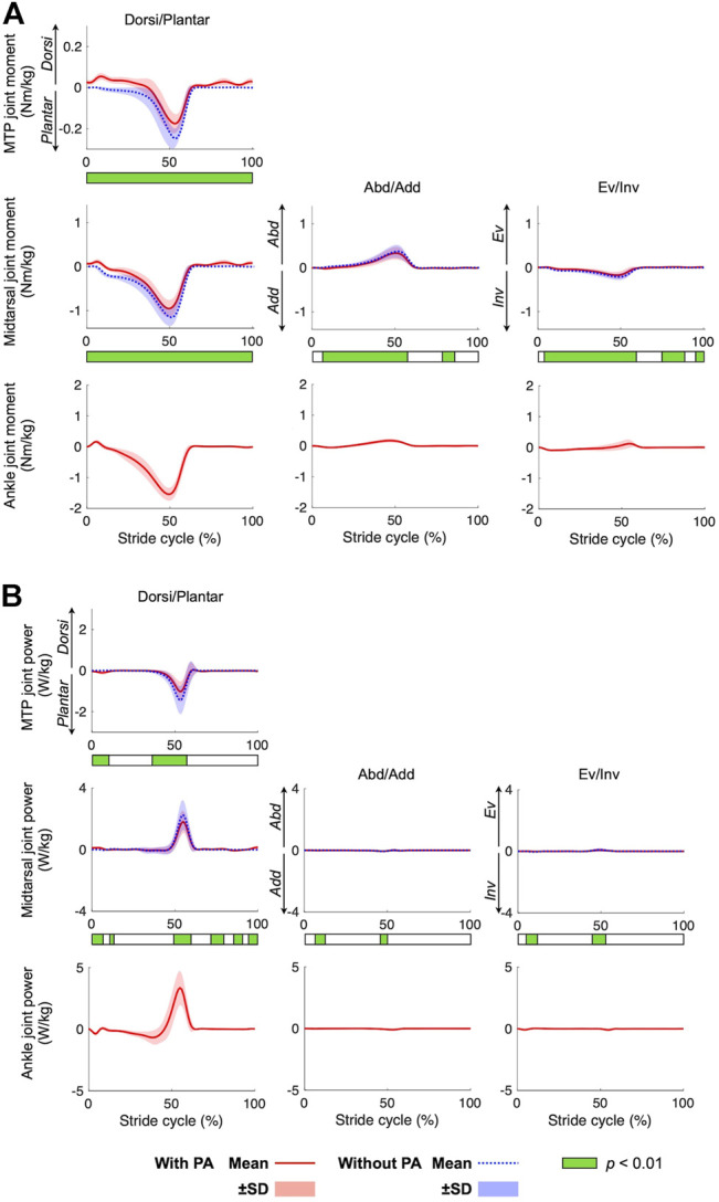
Kinetic multi-segment foot models have been proposed to evaluate the forces and moments generated in the foot during walking based on inverse dynamics calculations. However, these models did not consider the plantar aponeurosis (PA) despite its potential importance in generation of the ground reaction forces and storage and release of mechanical energy. This study aimed to develop a novel multi-segment foot model incorporating the PA to better elucidate foot kinetics. The foot model comprised three segments: the phalanx, forefoot, and hindfoot. The PA was modeled using five linear springs connecting the origins and the insertions via intermediate points. To demonstrate the efficacy of the foot model, an inverse dynamic analysis of human gait was performed and how the inclusion of the PA model altered the estimated joint moments was examined. Ten healthy men walked along a walkway with two force plates placed in series close together. The attempts in which the participant placed his fore- and hindfoot on the front and rear force plates, respectively, were selected for inverse dynamic analysis. The stiffness and the natural length of each PA spring remain largely uncertain. Therefore, a sensitivity analysis was conducted to evaluate how the estimated joint moments were altered by the changes in the two parameters within a range reported by previous studies. The present model incorporating the PA predicted that 13%-45% of plantarflexion in the metatarsophalangeal (MTP) joint and 8%-29% of plantarflexion in the midtarsal joints were generated by the PA at the time of push-off during walking. The midtarsal joint generated positive work, whereas the MTP joint generated negative work in the late stance phase. The positive and negative work done by the two joints decreased, indicating that the PA contributed towards transfer of the energy absorbed at the MTP joint to generate positive work at the midtarsal joint during walking. Although validation is limited due to the difficulty associated with direct measurement of the PA force in vivo, the proposed novel foot model may serve as a useful tool to clarify the function and mechanical effects of the PA and the foot during dynamic movements.
Keywords: foot kinematics; foot kinetics; healthy adults; inverse dynamics; motion analysis; multi-segment foot model; plantar fascia; walking.
Copyright © 2022 Matsumoto, Ogihara, Hanawa, Kokubun and Kanemura.
Conflict of interest statement
The authors declare that the research was conducted in the absence of any commercial or financial relationships that could be construed as a potential conflict of interest.
Figures


Similar articles
-
Kinetic coupling in distal foot joints during walking.J Foot Ankle Res. 2023 Jul 25;16(1):44. doi: 10.1186/s13047-023-00643-x. J Foot Ankle Res. 2023. PMID: 37488576 Free PMC article. Clinical Trial.
-
Partitioning ground reaction forces for multi-segment foot joint kinetics.Gait Posture. 2018 May;62:111-116. doi: 10.1016/j.gaitpost.2018.03.001. Epub 2018 Mar 6. Gait Posture. 2018. PMID: 29544155 Free PMC article.
-
Foot arch rigidity in walking: In vivo evidence for the contribution of metatarsophalangeal joint dorsiflexion.PLoS One. 2022 Sep 8;17(9):e0274141. doi: 10.1371/journal.pone.0274141. eCollection 2022. PLoS One. 2022. PMID: 36074770 Free PMC article.
-
Biomechanical effects of rocker shoes on plantar aponeurosis strain in patients with plantar fasciitis and healthy controls.PLoS One. 2019 Oct 10;14(10):e0222388. doi: 10.1371/journal.pone.0222388. eCollection 2019. PLoS One. 2019. PMID: 31600227 Free PMC article.
-
Contributions to the understanding of gait control.Dan Med J. 2014 Apr;61(4):B4823. Dan Med J. 2014. PMID: 24814597 Review.
Cited by
-
Sex differences in the kinematics and kinetics of the foot and plantar aponeurosis during drop-jump.Sci Rep. 2023 Aug 10;13(1):12957. doi: 10.1038/s41598-023-39682-6. Sci Rep. 2023. PMID: 37563188 Free PMC article.
-
Foot kinematics and kinetics data for different static foot posture collected using a multi-segment foot model.Sci Data. 2024 Nov 30;11(1):1307. doi: 10.1038/s41597-024-04166-3. Sci Data. 2024. PMID: 39616167 Free PMC article.
-
Direct visualization and measurement of the plantar aponeurosis behavior in foot arch deformation via the windlass mechanism.Clin Anat. 2025 Mar;38(2):116-126. doi: 10.1002/ca.24171. Epub 2024 Apr 20. Clin Anat. 2025. PMID: 38642017 Free PMC article.
References
LinkOut - more resources
Full Text Sources