Review
. 2022 Jun 23:13:909686.
doi: 10.3389/fpsyg.2022.909686. eCollection 2022.

Choosing Sides: Impact of Prismatic Adaptation on the Lateralization of the Attentional System

Affiliations
Review

Choosing Sides: Impact of Prismatic Adaptation on the Lateralization of the Attentional System

Stephanie Clarke et al. Front Psychol. .

Abstract

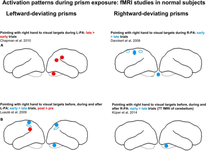
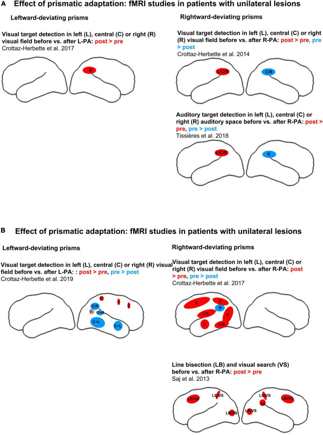
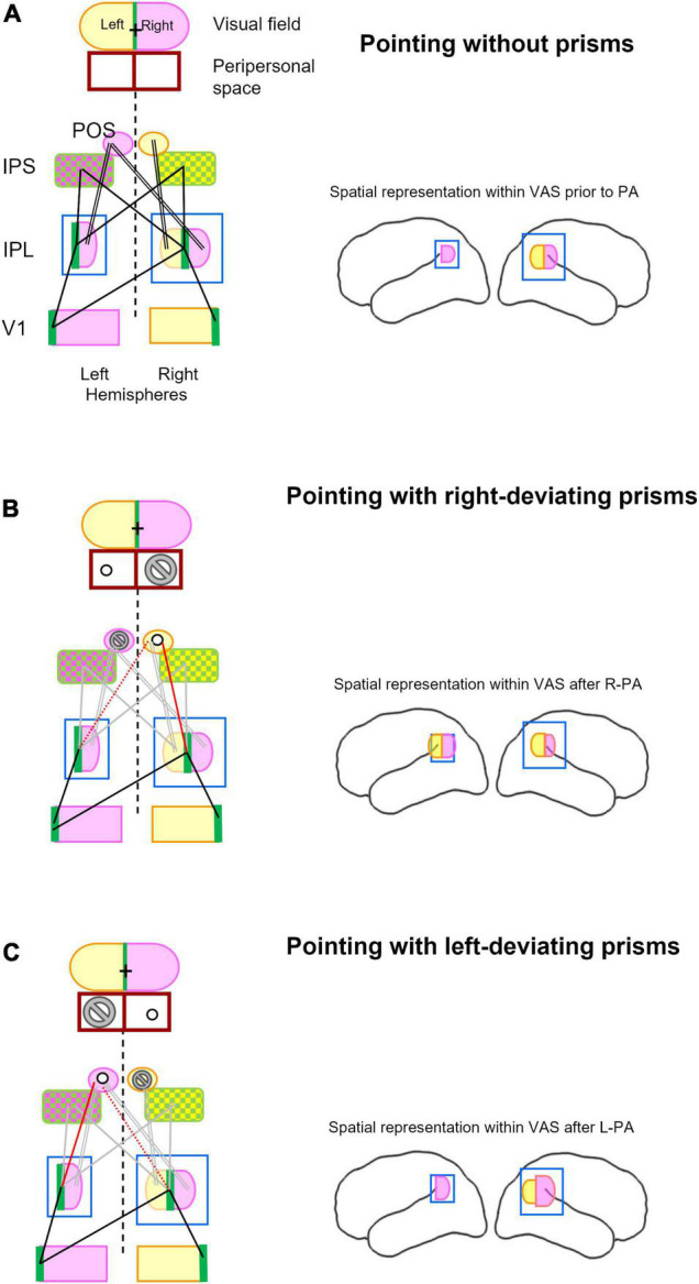
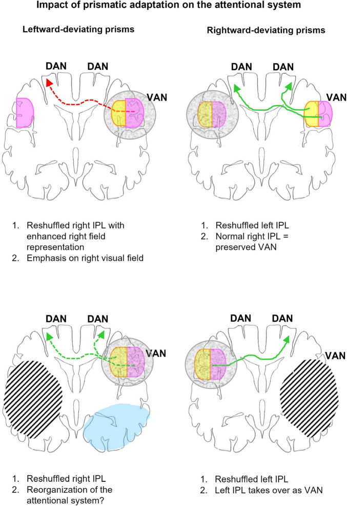
Seminal studies revealed differences between the effect of adaptation to left- vs. right-deviating prisms (L-PA, R-PA) in normal subjects. Whereas L-PA leads to neglect-like shift in attention, demonstrated in numerous visuo-spatial and cognitive tasks, R-PA has only minor effects in specific aspects of a few tasks. The paucity of R-PA effects in normal subjects contrasts with the striking alleviation of neglect symptoms in patients with right hemispheric lesions. Current evidence from activation studies in normal subjects highlights the contribution of regions involved in visuo-motor control during prism exposure and a reorganization of spatial representations within the ventral attentional network (VAN) after the adaptation. The latter depends on the orientation of prisms used. R-PA leads to enhancement of the ipsilateral visual and auditory space within the left inferior parietal lobule (IPL), switching thus the dominance of VAN from the right to the left hemisphere. L-PA leads to enhancement of the ipsilateral space in right IPL, emphasizing thus the right hemispheric dominance of VAN. Similar reshaping has been demonstrated in patients. We propose here a model, which offers a parsimonious explanation of the effect of L-PA and R-PA both in normal subjects and in patients with hemispheric lesions. The model posits that prismatic adaptation induces instability in the synaptic organization of the visuo-motor system, which spreads to the VAN. The effect is lateralized, depending on the side of prism deviation. Successful pointing with prisms implies reaching into the space contralateral, and not ipsilateral, to the direction of prism deviation. Thus, in the hemisphere contralateral to prism deviation, reach-related neural activity decreases, leading to instability of the synaptic organization, which induces a reshuffling of spatial representations in IPL. Although reshuffled spatial representations in IPL may be functionally relevant, they are most likely less efficient than regular representations and may thus cause partial dysfunction. The former explains, e.g., the alleviation of neglect symptoms after R-PA in patients with right hemispheric lesions, the latter the occurrence of neglect-like symptoms in normal subjects after L-PA. Thus, opting for R- vs. L-PA means choosing the side of major IPL reshuffling, which leads to its partial dysfunction in normal subjects and to recruitment of alternative or enhanced spatial representations in patients with hemispheric lesions.

Keywords: fMRI; prism-induced plasticity; review; spatial representation; ventral attentional network.


Conflict of interest statement

The authors declare that the research was conducted in the absence of any commercial or financial relationships that could be construed as a potential conflict of interest.

Figures

FIGURE 1
FIGURE 1
Effects of adaptation to left-deviating (left column) and right-deviating prisms (right column), as demonstrated in normal subjects with fMRI paradigms. (A) Changes of activity elicited by pointing during prism exposure. (B) Changes of activity elicited by pointing before, during, and after adaptation. Red and blue denote, respectively, increases and decreases of activity when comparing early vs. late stages of adaptation or pre- vs. post-adaptation sessions. Filled circles and ellipses mark cluster of activity on the convexity, empty ones those in sulci or on the medial part of the hemisphere.
FIGURE 2
FIGURE 2
Effects of adaptation to left-deviating (left column) or right-deviating prisms (right column), as demonstrated with fMRI paradigms. (A) Changes of activity elicited by visual target detection before vs. after prismatic adaptation in normal subjects. (B) Changes of activity elicited by visual target detection (top row) or by line bisection and visual search before vs. after prismatic adaptation (bottom row) in patients with left or right unilateral hemispheric lesions (left and right columns, respectively). Red and blue denote, respectively, increases and decreases of activity when comparing early vs. late stages of adaptation or pre- vs. post-adaptation sessions. Filled circles and ellipses mark cluster of activity on the convexity, empty ones those in sulci or on the medial part of the hemisphere. L, C, and R denote, respectively, left, central, and right stimulus position in target detection paradigms. LB denotes line bisection, VS visual search.
FIGURE 3
FIGURE 3
Schematic representation of key structures involved in visual pointing and their topographical relationship while pointing without prism (A), with right-deviating prisms (B), and with left-deviating prisms (C). Lateral views of the left and right hemispheres in right-hand column summarize spatial representation within the inferior parietal lobule and its changes after prismatic adaptation as described previously (Crottaz-Herbette et al., 2014, 2017b; Tissieres et al., 2018). (A) The visual field is subdivided into right (violet), left (yellow), and vertical meridian parts (green). Left and right halves of peripersonal space are outlined below the visual field figurine and are aligned with it, representing thus the situation of pointing to central (visual) targets. The same color code, violet for right, yellow for left, and green for central visual field, is used for the respective representations within the primary visual area (V1), the inferior parietal lobule (IPL), the intraparietal sulcus (IPS), and the parieto-occipital sulcus (POS). For simplicity reason, the extra-striate visual areas are not included. Whereas IPS encodes central (green) and contralateral targets (violet and yellow, respectively), POS encodes only the contralateral targets. Pointing to visual targets at (central) fixation is the most accurate (Prado et al., 2005) and most commonly adopted. Without prisms, pointing to central target involved central visual field representations (green) in visual areas (here represented by V1), IPL and IPS. Main topographic relationships involved in pointing to central targets are indicated here by single black lines, those involved in pointing to peripheral targets by double black lines. Brain figurine on the right depicts bilateral space representation in right IPL. (B) Pointing to visual targets while wearing right-deviating prisms implies creating a relationship between the visually perceived target at fixation point (cross in visual field; green in V1 and IPL) and the actual target in the peripersonal space to the left of the fixation point (circle). It is to be noted that the actual target will never be in the peripersonal space to the right of the fixation point (marked by the no-go sign). This new configuration weakens links between the central representations within IPL and IPS on either side (gray lines) and between right space representation within right and left IPL and the left IPS and POS (double lines in gray). It will establish a new link between the central representation in IPL on the right (and possibly the left) side and the left space representation within right IPS and POS (red line), possibly by re-adjusting a previous link between the left space in these structures. Brain figurine on the right depicts R-PA-induced bilateral space representation in left IPL (Crottaz-Herbette et al., 2014; Tissieres et al., 2018). (C) Pointing to visual targets while wearing left-deviating prisms implies creating a relationship between the visually perceived target at fixation point (cross in visual field; green in V1 and IPL) and the actual target in the peripersonal space to the right of the fixation point (circle). It is to be noted that the actual target will never be in the peripersonal space to the left of the fixation point (marked by the no-go sign). This new configuration weakens links between the central representations within IPL and IPS on either side (gray lines) and between left space representation within right IPL and right IPS and POS (double line in gray). It will establish a new link between the central representation in IPL on the left (and possibly right) side and the right space representation within the left IPS and POS (red line), possibly by re-adjusting a previous link between the right space in these structures (dotted red line). Brain figurine on the right depicts L-PA-induced reinforcement of right space representation in right IPL (Crottaz-Herbette et al., 2017b).
FIGURE 4
FIGURE 4
Schematic representation of the impact of adaptation to left- (left column) and right-deviating prisms (right column) on the organization of the attentional network in normal subjects (top row) and patients with unilateral hemispheric lesions (bottom row). The outline represents a coronal section at the level of IPL, right hemisphere is to the right. Gray mottled overlay marks reshuffled IPL representations. Right space representation is in violet, left space in yellow. Green arrows indicate links relevant to correct or recovered function, red arrows those contributing to dysfunction. Arrows in full line mark links compatible with behavioral and/or imaging evidence, those in dashed line, hypotheses to be tested. Hatching represents lesions, blue, regions where L-PA downregulates activation by visual stimuli. Top: In normal subjects L-PA leads to the reshuffling of spatial representations within VAN, enhancing the ipsilateral space representation (Crottaz-Herbette et al., 2017b). The latter is likely to increase the activation of DAN in the left hemisphere. Both the relative dysfunction of the reshuffled VAN and increased activation of left DAN are likely to contribute to the neglect-like effects, which L-PA induces in normal subjects. R-PA leads in normal subjects to the reshuffling of spatial representations within left IPL, by enhancing the ipsilateral space representation (Crottaz-Herbette et al., 2014; Tissieres et al., 2018). Since VAN is preserved, the reshuffled left IPL is very likely not to impact attentional functions. Bottom: In patients with right-hemispheric lesions, R-PA leads to the reshuffling of spatial representations within left IPL, by enhancing the ipsilateral space representation (Crottaz-Herbette et al., 2017a). In the absence of a functional VAN, the left IPL takes over and drives DAN on either side. In patients with left-hemispheric lesions, L-PA reshuffles and changes partially spatial representations within VAN; in addition it downregulates visual activity in the extrastriate cortex (Crottaz-Herbette et al., 2019). The reorganization of VAN may have a positive impact on attentional functioning.

Similar articles

Cited by

References

    1. Baizer J. S., Kralj-Hans I., Glickstein M. (1999). Cerebellar lesions and prism adaptation in macaque monkeys. J. Neurophysiol. 81 1960–1965. 10.1152/jn.1999.81.4.1960 - DOI - PubMed
    1. Barrett A. M., Goedert K. M., Basso J. C. (2012). Prism adaptation for spatial neglect after stroke: translational practice gaps. Nat. Rev. Neurol. 8 567–577. 10.1038/nrneurol.2012.170 - DOI - PMC - PubMed
    1. Barrett A. M., Houston K. E. (2019). Update on the clinical approach to spatial neglect. Curr. Neurol. Neurosci. Rep. 19:25. 10.1007/s11910-019-0940-0 - DOI - PMC - PubMed
    1. Berberovic N., Mattingley J. B. (2003). Effects of prismatic adaptation on judgements of spatial extent in peripersonal and extrapersonal space. Neuropsychologia 41 493–503. 10.1016/S0028-3932(02)00090-8 - DOI - PubMed
    1. Beume L.-A., Kaller C. P., Hoeren M., Klöppel S., Kuemmerer D., Glauche V., et al. (2015). Processing of bilateral versus unilateral conditions: evidence for the functional contribution of the ventral attention network. Cortex 66 91–102. 10.1016/j.cortex.2015.02.018 - DOI - PubMed