

Shuangxinfang Prevents S100A9-Induced Macrophage/Microglial Inflammation to Improve Cardiac Function and Depression-Like Behavior in Rats After Acute Myocardial Infarction
- PMID: 35814253
- PMCID: PMC9263923
- DOI: 10.3389/fphar.2022.832590
Shuangxinfang Prevents S100A9-Induced Macrophage/Microglial Inflammation to Improve Cardiac Function and Depression-Like Behavior in Rats After Acute Myocardial Infarction
Abstract
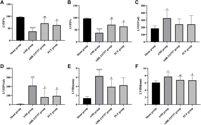
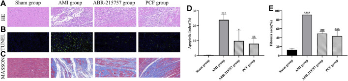
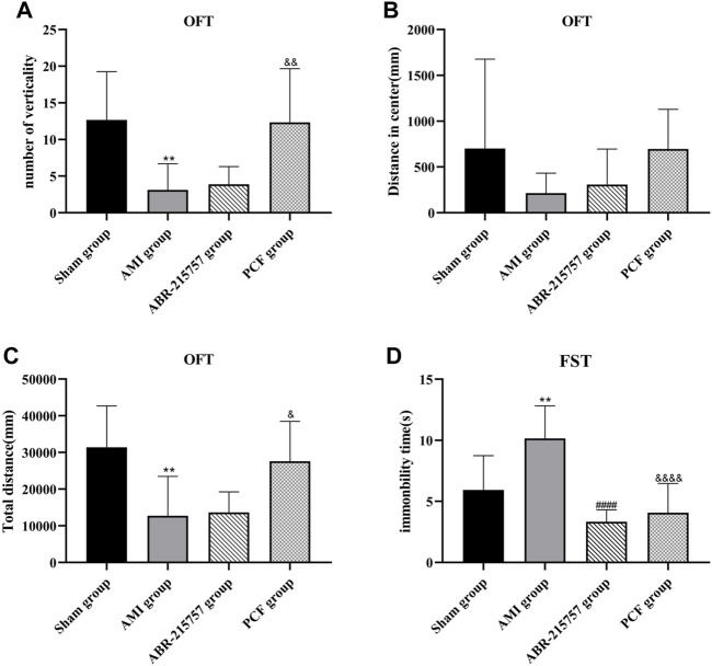
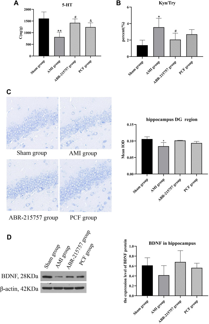
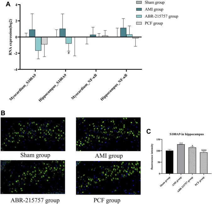
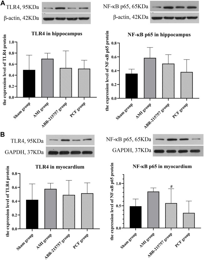
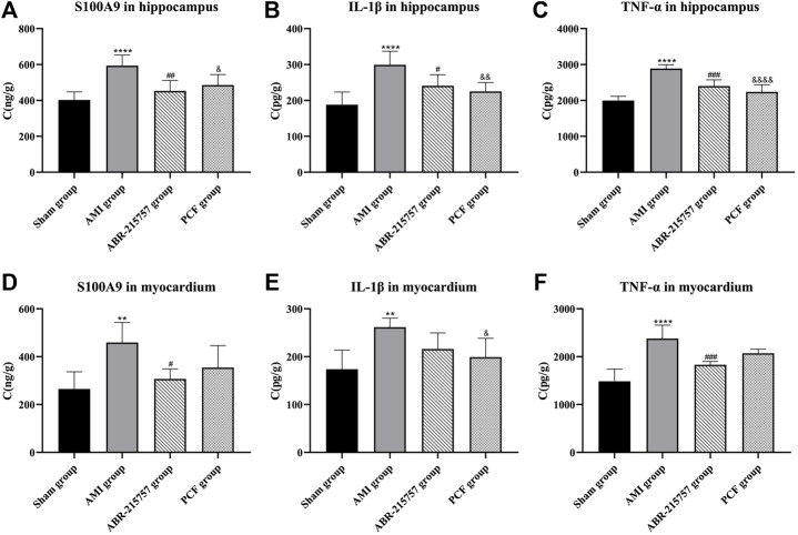
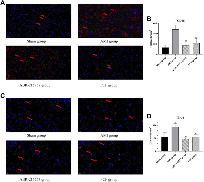
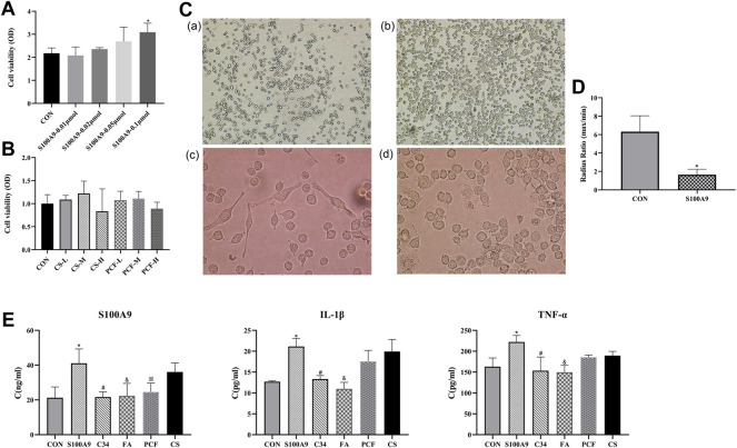
Background: Depression is a common complication of cardiovascular disease, which deteriorates cardiac function. Shuangxinfang (psycho-cardiology formula, PCF) was reported to alleviate myocardial ischemia injury and improve depression-like behavior. Interestingly, our previous proteomics study predicted that the protein S100A9 appeared as an important target, and macrophage/microglial inflammation might be involved in the process of PCF improving depression induced by acute myocardial infarction (AMI). This study aims to validate the proteomics results. Methods: AMI rat models were established in vivo, followed by the administration of PCF or ABR-215757 (also named paquinimod, inhibiting S100A9 binding to TLR4) for 5 days. Forced swimming test (FST) and open field test (OFT) were applied to record depression-like behavior, and echocardiography was employed to evaluate cardiac function. Morphological changes of cardiomyocytes were assessed by HE staining and TUNEL staining on day 7 after cardiac surgery, as well as Masson trichrome staining on day 21. Hippocampal neurogenesis was determined by Nissl staining, while 5-hydroxytryptamine (5-HT), tryptophan/kynurenine ratio, and brain-derived neurotrophic factor (BDNF) in the hippocampus were analyzed as biochemical indicators of depression. We employed RT-qPCR, western blotting, and immunofluorescence to detect the expression of pathway-related genes and proteins. Myocardial and hippocampal expression of inflammatory factors were performed by ELISA. The activation of macrophage and microglia was assessed via immunoreaction using CD68 and Iba1, respectively. For in vitro confirmation, BV2 cells were primed with recombinant protein S100A9 and then treated with PCF serum or ferulic acid to determine alterations in microglial inflammation. Results: Rats in the AMI group showed heart function deterioration and depression-like behavior. Coronary ligation not only brought about myocardial inflammation, cell apoptosis, and fibrosis but also reduced the neurogenesis, elevated the tryptophan/kynurenine ratio, and decreased the content of 5-HT. PCF could ameliorate the pathological and phenotypic changes in the heart and brain and inhibit the expression of the S100A9 protein, the activation of the microglial cell, and the secretion of IL-1β and TNF-α raised by AMI. ABR-215757 showed therapeutic effect and molecular biological mechanisms similar to PCF. Treatment with PCF serum or ferulic acid in vitro was proved to efficiently block the hyperactivation of BV2 cells and increment of cytokine contents induced by recombinant protein S100A9. Conclusion: We identify S100A9 as a novel and potent regulator of inflammation in both the heart and brain. Macrophage/microglia inflammation mediated by S100A9 is considered a pivotal pathogenic in depression after AMI and a major pathway for the treatment of PCF, suggesting that PCF is a promising therapeutic candidate for psycho-cardiology disease.
Keywords: S100A9; Shuangxinfang; acute myocardial infarction; epressive disorder; inflammation; macrophages; microglia; traditional Chinese medicine.
Copyright © 2022 Sun, Wang, Hou, Shi, Tang, Wang and Zhao.
Conflict of interest statement
The authors declare that the research was conducted in the absence of any commercial or financial relationships that could be construed as a potential conflict of interest.
Figures
Similar articles
-
Psycho-cardiology therapeutic effects of Shuangxinfang in rats with depression-behavior post acute myocardial infarction: Focus on protein S100A9 from proteomics.Biomed Pharmacother. 2021 Dec;144:112303. doi: 10.1016/j.biopha.2021.112303. Epub 2021 Oct 19. Biomed Pharmacother. 2021. PMID: 34673424
-
Compound identification of Shuangxinfang and its potential mechanisms in the treatment of myocardial infarction with depression: insights from LC-MS/MS and bioinformatic prediction.Front Pharmacol. 2025 Jan 28;16:1499418. doi: 10.3389/fphar.2025.1499418. eCollection 2025. Front Pharmacol. 2025. PMID: 39936089 Free PMC article.
-
Astaxanthin promotes M2 macrophages and attenuates cardiac remodeling after myocardial infarction by suppression inflammation in rats.Chin Med J (Engl). 2020 Aug 5;133(15):1786-1797. doi: 10.1097/CM9.0000000000000814. Chin Med J (Engl). 2020. PMID: 32701588 Free PMC article.
-
Inflammatory Mediators in Pericardial Fluid in Patients Undergoing Cardiac Surgery.CJC Open. 2024 Nov 8;7(2):193-202. doi: 10.1016/j.cjco.2024.11.001. eCollection 2025 Feb. CJC Open. 2024. PMID: 40060216 Free PMC article. Review.
-
Cardiovascular disease and depression: a narrative review.Front Cardiovasc Med. 2023 Nov 21;10:1274595. doi: 10.3389/fcvm.2023.1274595. eCollection 2023. Front Cardiovasc Med. 2023. PMID: 38084332 Free PMC article. Review.
Cited by
-
Honokiol relieves hippocampal neuronal damage in Alzheimer's disease by activating the SIRT3-mediated mitochondrial autophagy.CNS Neurosci Ther. 2024 Aug;30(8):e14878. doi: 10.1111/cns.14878. CNS Neurosci Ther. 2024. PMID: 39097923 Free PMC article.
-
Effects of the LINC00641/miR-323a-3p/EIF4G2 axis on behaviors and brain monoamine neurotransmitters in chronic unpredictable mild stress mice.Cell Biol Toxicol. 2025 Apr 28;41(1):76. doi: 10.1007/s10565-025-10015-9. Cell Biol Toxicol. 2025. PMID: 40293545 Free PMC article.
-
Identification and validation of lipid metabolism-related key genes as novel biomarkers in acute myocardial infarction and pan-cancer analysis.Aging (Albany NY). 2024 May 23;16(10):9127-9146. doi: 10.18632/aging.205860. Epub 2024 May 23. Aging (Albany NY). 2024. PMID: 38787365 Free PMC article.
-
Advances in the study of S100A9 in cardiovascular diseases.Cell Prolif. 2024 Aug;57(8):e13636. doi: 10.1111/cpr.13636. Epub 2024 Mar 19. Cell Prolif. 2024. PMID: 38504474 Free PMC article. Review.
-
Recent advances of traditional Chinese medicine against cardiovascular disease: overview and potential mechanisms.Front Endocrinol (Lausanne). 2024 Sep 30;15:1366285. doi: 10.3389/fendo.2024.1366285. eCollection 2024. Front Endocrinol (Lausanne). 2024. PMID: 39403576 Free PMC article. Review.
References
-
- Bah T. M., Benderdour M., Kaloustian S., Karam R., Rousseau G., Godbout R. (2011a). Escitalopram Reduces Circulating Pro-inflammatory Cytokines and Improves Depressive Behavior without Affecting Sleep in a Rat Model of Post-cardiac Infarct Depression. Behav. Brain Res. 225 (1), 243–251. 10.1016/j.bbr.2011.07.039 - DOI - PubMed
-
- Bangalore S., Shah R., Gao X., Pappadopulos E., Deshpande C. G., Shelbaya A., et al. (2020). Economic Burden Associated with Inadequate Antidepressant Medication Management Among Patients with Depression and Known Cardiovascular Diseases: Insights from a United States-based Retrospective Claims Database Analysis. J. Med. Econ. 23 (3), 262–270. 10.1080/13696998.2019.1686311 - DOI - PubMed
LinkOut - more resources
Full Text Sources
Miscellaneous