. 2022 Jun 29:2022:2948090.
doi: 10.1155/2022/2948090. eCollection 2022.

circ_0072464 Shuttled by Bone Mesenchymal Stem Cell-Secreted Extracellular Vesicles Inhibits Nucleus Pulposus Cell Ferroptosis to Relieve Intervertebral Disc Degeneration

Affiliations

circ_0072464 Shuttled by Bone Mesenchymal Stem Cell-Secreted Extracellular Vesicles Inhibits Nucleus Pulposus Cell Ferroptosis to Relieve Intervertebral Disc Degeneration

Xiaojun Yu et al. Oxid Med Cell Longev. .

Abstract

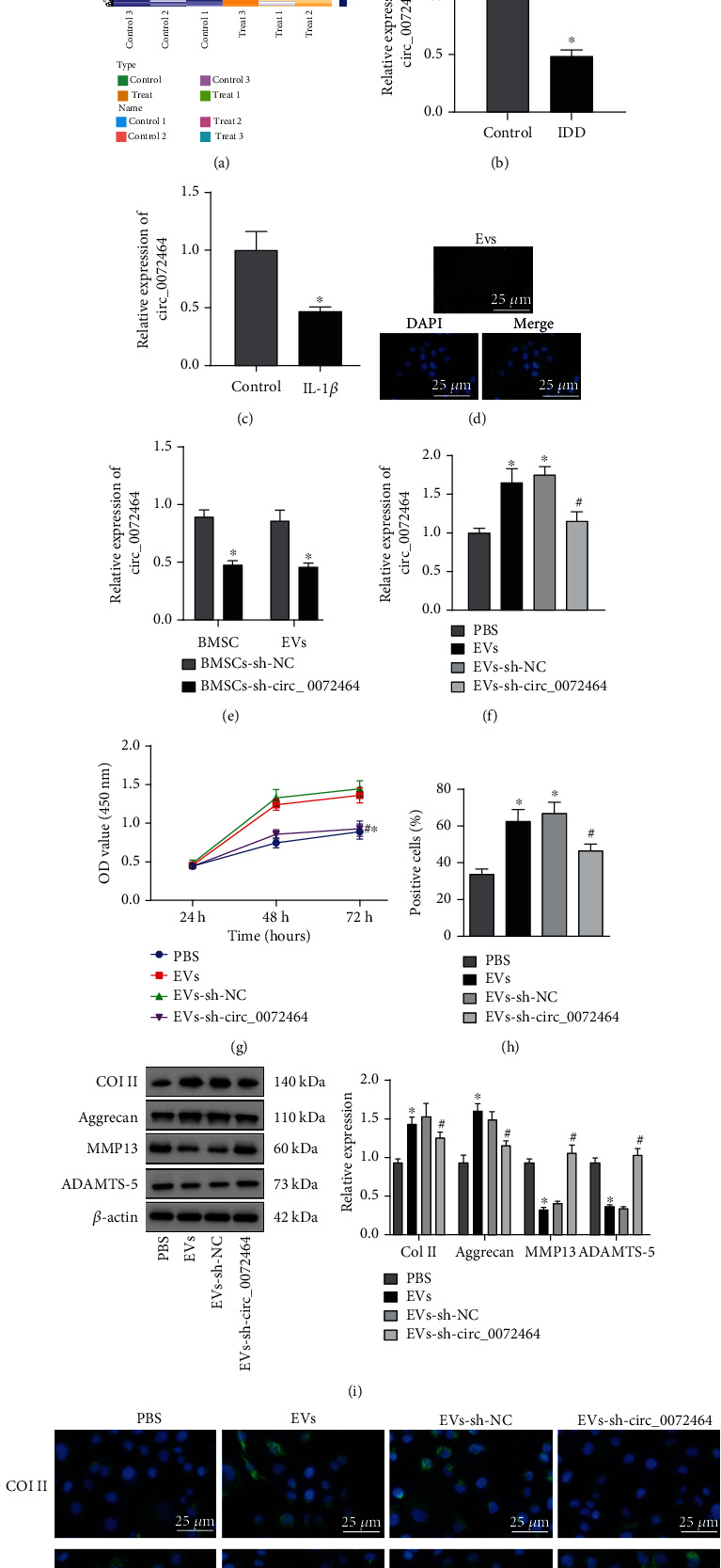
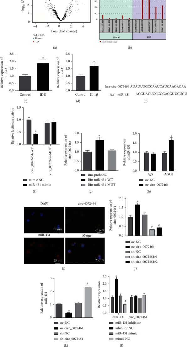
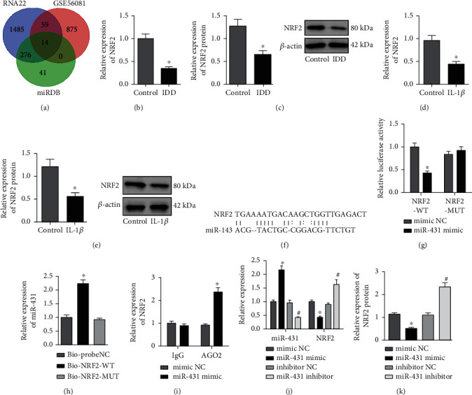
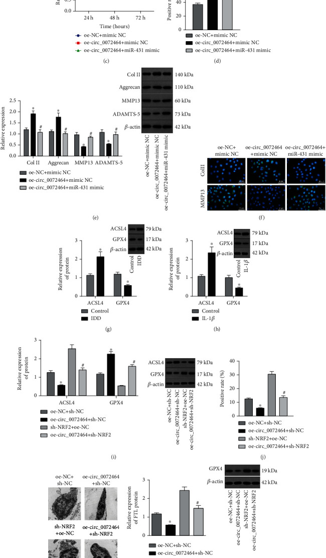
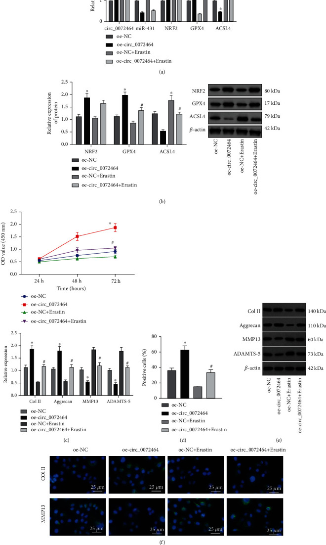
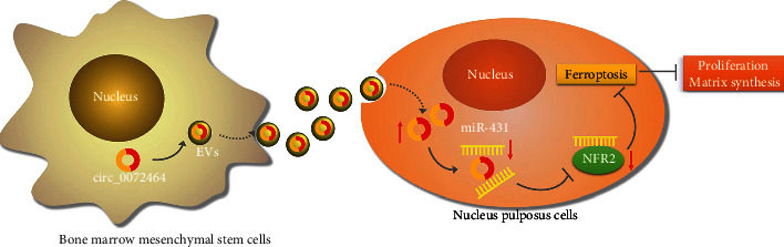
Ferroptosis, as an iron-dependent form of necrotic cell death, has been reported to affect activities of nucleus pulposus cells (NPCs). However, its role in the pathogenesis of intervertebral disc degeneration (IDD) is largely unknown. Notably, our bioinformatics analysis predicted that circ_0072464 was downregulated in nucleus pulposus of IDD mice. Therefore, this study is aimed at clarifying the mechanisms of extracellular vesicle- (EV-) encapsulated circ_0072464 derived from bone marrow mesenchymal stem cells (BMSCs) in NPC ferroptosis in IDD. EVs were extracted from mouse BMSCs (BMSC-EVs) and then cocultured with IL-1β-induced NPCs, followed by detection of matrix synthesis, proliferation, and ferroptosis of NPCs based on gain- or loss-of-function experiments. It was found that the uptake of BMSC-EVs by NPCs alleviated IDD. circ_0072464 and NRF2 were downregulated, and miR-431 was upregulated in IDD. Mechanistically, circ_0072464 competitively bound to miR-431, which targeted and inhibited NRF2 expression. BMSC-derived EVs carrying circ_0072464 inhibited NPC ferroptosis to promote matrix synthesis and proliferation of NPCs by inhibiting miR-431 and upregulating NRF2. Besides, in vivo experiments also confirmed that BMSC-EVs alleviated intervertebral disc lesions in mice with IDD through the circ_0072464/miR-431/NRF2 axis. Collectively, BMSC-EV-loaded circ_0072464 inhibited NPC ferroptosis to relieve IDD via upregulation of miR-431-mediated NRF2, therefore providing a potential therapeutic target against IDD.


Conflict of interest statement

The authors have declared that no competing interest exists.

Figures

Figure 1
Figure 1
Effects of BMSC-EVs on IDD mice. (a) The morphology of EVs observed under the TEM. (b) The particle size distribution of EVs analyzed by NTA. (c) The protein levels of EV markers CD63, TSG101, and CD81 and negative control calnexin detected by Western blot analysis. (d) The NP and cartilage AF of IVD after modeling detected by safranin O staining (n = 10). The first row shows the staining image of IVD as a whole, the second row displays the detailed view of NP tissues, and the third row presents the detailed view of boundary between NP and AF tissues. p < 0.05vs. IDD mice at the 6th month (M). (e) The histopathology of NP and the AF of NP and cartilage detected by safranin O staining (n = 10). The first row shows the staining image of IVD as a whole, the second row displays the detailed view of NP tissues, and the third row presents the detailed view of boundary between NP and AF tissues. p < 0.05vs. IDD mice and #p < 0.05vs. IDD mice at the 6th week (W). (f) The levels of Col II and MMP13 in NP tissues measured by immunofluorescence staining (n = 10). Cell experiments were repeated for three times independently.
Figure 2
Figure 2
circ_0072464 shuttled by BMSC-EVs affects matrix synthesis and proliferation of NPCs. (a) Heat map of differentially expressed circRNAs screened from RNA-seq data (GSE189551) of NP tissues of IDD mice and normal mice. (b) circ_0072464 expression in NP tissues of IDD mice and normal mice (n = 10, p < 0.05vs. control mice). (c) circ_0072464 expression in NPCs determined by RT-qPCR (p < 0.05vs. control NPCs). (d) The uptake of EVs by NPCs observed under the confocal microscope; blue: Hoechst staining; green: PKH67-labeled EVs. (e) circ_0072464 expression in BMSC-EVs in response to sh-circ_0072464 determined by RT-qPCR (p < 0.05vs. BMSCs transduced with sh-NC). (f) circ_0072464 expression in NPCs after coculture with EVs or EVs-sh-circ_0072464 determined by RT-qPCR. (g) Viability of NPCs after coculture with EVs or EVs-sh-circ_0072464 detected by CCK8 assay. (h) Proliferation of NPCs after coculture with EVs or EVs-sh-circ_0072464 detected by EdU assay. (i) Protein levels of Col II, Aggrecan, MMP13, and ADAMTS-5 in NPCs after coculture with EVs or EVs-sh-circ_0072464 measured by Western blot analysis. (j) Expression of Col II and MMP13 in NPCs after coculture with EVs or EVs-sh-circ_0072464 determined by immunofluorescence staining. In (f–j), p < 0.05vs. PBS-treated NPCs and #p < 0.05vs. NPCs cocultured with EVs-sh-NC. Cell experiments were repeated for three times independently.
Figure 3
Figure 3
circ_0072464 sponges miR-431. (a) miR-431 expression in IDD sample of GSE63492 dataset. (b) Volcano map of differential analysis for GSE63492 dataset. (c) miR-431 expression in NP tissues of IDD mice determined by RT-qPCR (n = 10, p < 0.05vs. control mice). (d) miR-431 expression in IL-1β-treated NPCs determined by RT-qPCR (p < 0.05vs. control NPCs). (e) The binding site between circ_0072464 and miR-431 predicted by bioinformatics website. (f) The relationship between circ_0072464 and miR-431 verified by dual-luciferase reporter gene assay (p < 0.05vs. mimic NC). (g) The interaction between circ_0072464 and miR-431 detected by RNA pull-down assay (p < 0.05vs. Bioprobe NC or Bio-miR-431-MUT). (h) The interaction between circ_0072464 and miR-431 detected by RIP assay (p < 0.05vs. oe-NC). (i) The colocalization between circ_0072464 and miR-431 in NPCs detected by RNA FISH assay. (j) circ_0072464 expression in NPCs in response to oe-circ_0072464 or sh-circ_0072464 determined by RT-qPCR (p < 0.05vs. NPCs transduced with oe-NC; #p < 0.05vs. NPCs transduced with sh-NC). (k) miR-431 expression in NPCs in response to oe-circ_0072464 or sh-circ_0072464 determined by RT-qPCR (p < 0.05vs. NPCs transduced with oe-NC; #p < 0.05vs. NPCs transduced with sh-NC). (l) Expression of circ_0072464 and miR-431 in NPCs in response to miR-431 inhibitor or mimic determined by RT-qPCR (p < 0.05vs. NPCs transfected with mimic NC; #p < 0.05vs. NPCs transfected with inhibitor NC). Cell experiments were repeated for three times independently.
Figure 4
Figure 4
miR-431 targets and negatively regulates NRF2. (a) Prediction of the downstream target genes of miR-431. The green circle represents the downstream target gene predicted by miRDB database. The blue circle represents the downstream target gene predicted by RNA22 database. The red circle represents the significantly downregulated mRNA screened in the IDD sample of GSE56081 dataset. The middle part represents the intersection of three datasets. (b) NRF2 mRNA level in NP tissues of IDD mice determined by RT-qPCR (n = 10, p < 0.05vs. control mice). (c) NRF2 protein level in NP tissues of IDD mice determined by Western blot analysis (n = 10, p < 0.05vs. control mice). (d) NRF2 mRNA level in IL-1β-treated NPCs determined by RT-qPCR (p < 0.05vs. control NPCs). (e) NRF2 protein level in IL-1β-treated NPCs determined by Western blot analysis (p < 0.05vs. control NPCs). (f) The binding site between miR-431 and NRF2 predicted by miRDB database. (g) The relationship between miR-431 and NRF2 verified by dual-luciferase reporter gene assay (p < 0.05vs. mimic NC). (h) The relationship between miR-431 and NRF2 verified by RNA pull-down assay (p < 0.05vs. Bioprobe NC or Bio-NRF2-MUT). (i) The interaction between miR-431 and NRF2 detected by RIP assay (p < 0.05vs. mimic NC). (j) miR-431 expression and NRF2 mRNA level in NPCs in response to miR-431 mimic or miR-431 inhibitor determined by RT-qPCR (p < 0.05vs. NPCs transfected with mimic NC; #p < 0.05vs. NPCs transfected with inhibitor NC). (k) NRF2 protein level in NPCs in response to miR-431 mimic or miR-431 inhibitor determined by Western blot analysis (p < 0.05vs. NPCs transfected with mimic NC; #p < 0.05vs. NPCs transfected with inhibitor NC). Cell experiments were repeated for three times independently.
Figure 5
Figure 5
circ_0072464 affects NPC ferroptosis via the miR-431/NRF2 axis. (a) Expression of circ_0072464, miR-431, and NRF2 in NPCs transduced with oe-circ_0072464 alone or combined with miR-431 mimic determined by RT-qPCR (p < 0.05vs. NPCs transduced with oe-NC+mimic NC and #p < 0.05vs. NPCs transduced with oe-circ_0072464+mimic NC). (b) NRF2 protein level in NPCs transduced with oe-circ_0072464 alone or combined with miR-431 mimic determined by Western blot analysis (p < 0.05vs. NPCs transduced with oe-NC+mimic NC and #p < 0.05vs. NPCs transduced with oe-circ_0072464+mimic NC). (c) Viability of NPCs transduced with oe-circ_0072464 alone or combined with miR-431 mimic detected by CCK8 assay (p < 0.05vs. NPCs transduced with oe-NC+mimic NC and #p < 0.05vs. NPCs transduced with oe-circ_0072464+mimic NC). (d) Proliferation of NPCs transduced with oe-circ_0072464 alone or combined with miR-431 mimic detected by EdU assay (p < 0.05vs. NPCs transduced with oe-NC+mimic NC and #p < 0.05vs. NPCs transduced with oe-circ_0072464+mimic NC). (e) Protein levels of Col II, Aggrecan, MMP13, and ADAMTS-5 in NPCs transduced with oe-circ_0072464 alone or combined with miR-431 mimic measured by Western blot analysis (p < 0.05vs. NPCs transduced with oe-NC+mimic NC and #p < 0.05vs. NPCs transduced with oe-circ_0072464+mimic NC). (f) Expression of Col II and MMP13 in NPCs transduced with oe-circ_0072464 alone or combined with miR-431 mimic determined by immunofluorescence staining. (g) Protein levels of GPX4 and ACSL4 in NP tissues of IDD mice determined by Western blot analysis (n = 10, p < 0.05vs. control mice). (h) Protein levels of GPX4 and ACSL4 in IL-1β-treated NPCs determined by Western blot analysis (p < 0.05vs. control NPCs). (i) Protein levels of GPX4 and ACSL4 in NPCs transduced with oe-circ_0072464 and/or sh-NRF2 determined by Western blot analysis. (j) Cellular lipid ROS level of NPCs transduced with oe-circ_0072464 and/or sh-NRF2 detected by flow cytometry. (k) Mitochondrial morphology of NPCs transduced with oe-circ_0072464 and/or sh-NRF2 observed under the TEM. (l) FTL protein level in NPCs transduced with oe-circ_0072464 and/or sh-NRF2 measured by Western blot analysis. (m) Measurement of free iron levels in NPCs transduced with oe-circ_0072464 and/or sh-NRF2. In (e–i), p < 0.05vs. NPCs transduced with oe-NC+sh-NC; #p < 0.05vs. NPCs transduced with oe-circ_0072464+sh-NC. Cell experiments were repeated for three times independently.
Figure 6
Figure 6
circ_0072464/miR-431/NRF2 affects matrix synthesis and proliferation of NPCs by regulating NPC ferroptosis. (a) Expression of circ_0072464, miR-431, NRF2, GPX4, and ACSL4 in NPCs in response to oe-circ_0072464 and/or Erastin determined by RT-qPCR. (b) Protein levels of NRF2, GPX4, and ACSL4 in NPCs in response to oe-circ_0072464 and/or Erastin determined by Western blot analysis. (c) NPC proliferation detected by CCK8 assay. (d) NPC proliferation in response to oe-circ_0072464 and/or Erastin detected by EdU assay. (e) Protein levels of Col II, Aggrecan, MMP13, and ADAMTS-5 in NPCs in response to oe-circ_0072464 and/or Erastin determined by Western blot analysis. (f) Expression of Col II, Aggrecan, MMP13, and ADAMTS-5 in NPCs in response to oe-circ_0072464 and/or Erastin determined by immunofluorescence staining. p < 0.05vs. NPCs transduced with oe-NC; #p < 0.05vs. NPCs transduced with oe-circ_0072464. Cell experiments were repeated for three times independently.
Figure 7
Figure 7
BMSC-EV-packaged circ_0072464 regulates miR-431/NRF2 to affect NPC ferroptosis in IDD mice. IDD mice were injected with EVs and/or Erastin (n = 10). (a) Expression of circ_0072464, miR-431, NRF2, GPX4, and ACSL4 in NP tissues of IDD mice measured by RT-qPCR. (b) Expression of NRF2, GPX4, and ACSL4 in NP tissues of IDD mice detected by immunohistochemistry, and expression of circ_0072464 (green fluorescence) and miR-431 (green fluorescence) in NP tissues of IDD mice detected by RNA FISH. (c) Protein levels of NRF2, GPX4, and ACSL4 in NP tissues of IDD mice determined by Western blot analysis. (d) Pathological changes of IVD, NP, and AF in IDD mice detected by safranin O staining. The first row shows the staining image of IVD as a whole, the second row displays the detailed view of NP tissues, and the third row presents the detailed view of boundary between NP and AF tissues. (e) Degeneration of IVD in IDD mice observed using X-ray. (f) Levels of Col II and MMP13 in NP tissues of IDD mice detected by immunofluorescence staining. p < 0.05vs. IDD mice injected with PBS; #p < 0.05vs. IDD mice injected with EVs.
Figure 8
Figure 8
Molecular mechanism of the effects of circ_0072464 shuttled by BMSCs-EVs on IDD by regulating NPC ferroptosis via miR-431/NRF2. circ_0072464 shuttled by BMSCs-EVs upregulates NRF2 expression by competitively binding to miR-431, thereby inhibiting ferroptosis and promoting matrix synthesis and proliferation of NPCs, ultimately relieving IDD.

Similar articles

Cited by

References

    1. Yang S., Zhang F., Ma J., Ding W. Intervertebral disc ageing and degeneration: the antiapoptotic effect of oestrogen. Ageing Research Reviews . 2020;57, article 100978 doi: 10.1016/j.arr.2019.100978. - DOI - PubMed
    1. Priyadarshani P., Li Y., Yao L. Advances in biological therapy for nucleus pulposus regeneration. Osteoarthritis and Cartilage . 2016;24(2):206–212. doi: 10.1016/j.joca.2015.08.014. - DOI - PubMed
    1. Desmoulin G. T., Pradhan V., Milner T. E. Mechanical aspects of intervertebral disc injury and implications on biomechanics. Spine . 2020;45(8):E457–E464. doi: 10.1097/BRS.0000000000003291. - DOI - PubMed
    1. Lu S., Song Y., Luo R., et al. Ferroportin-dependent iron homeostasis protects against oxidative stress-induced nucleus pulposus cell ferroptosis and ameliorates intervertebral disc degeneration in vivo. Oxidative Medicine and Cellular Longevity . 2021;2021:18. doi: 10.1155/2021/6670497.6670497 - DOI - PMC - PubMed
    1. Zhang X., Huang Z., Xie Z., et al. Homocysteine induces oxidative stress and ferroptosis of nucleus pulposus via enhancing methylation of GPX4. Free Radical Biology & Medicine . 2020;160:552–565. doi: 10.1016/j.freeradbiomed.2020.08.029. - DOI - PubMed

MeSH terms

LinkOut - more resources