

Extracellular Vesicles and Resistance to Anticancer Drugs: A Tumor Skeleton Key for Unhinging Chemotherapies
- PMID: 35814444
- PMCID: PMC9259994
- DOI: 10.3389/fonc.2022.933675
Extracellular Vesicles and Resistance to Anticancer Drugs: A Tumor Skeleton Key for Unhinging Chemotherapies
Abstract
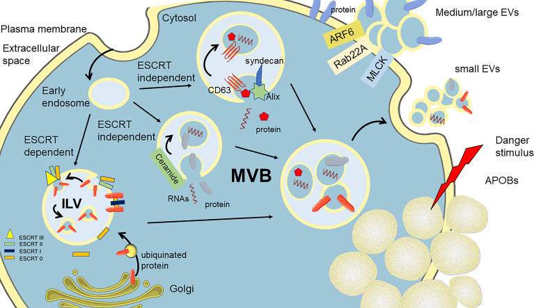
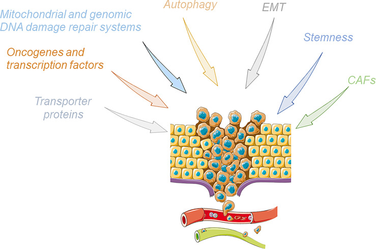
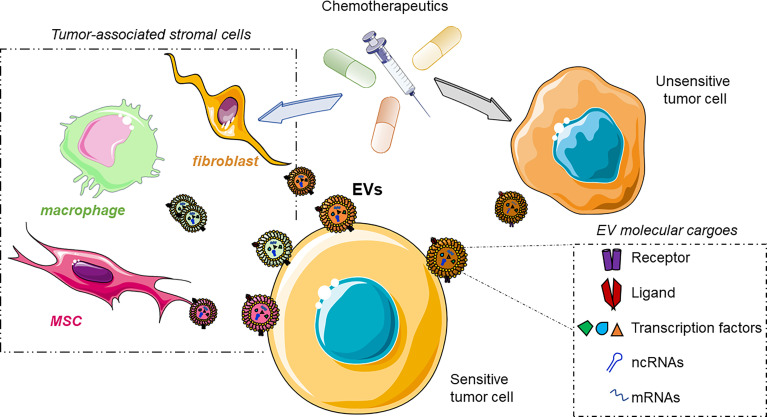
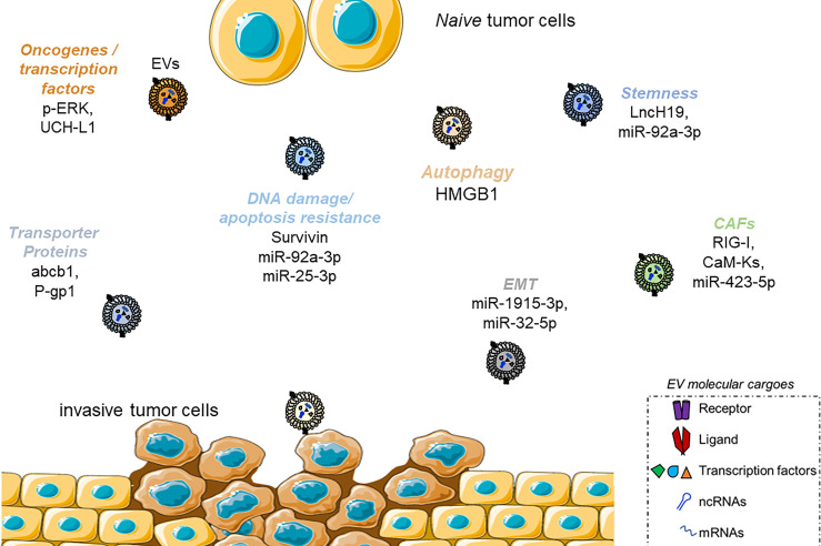
Although surgical procedures and clinical care allow reaching high success in fighting most tumors, cancer is still a formidable foe. Recurrence and metastatization dampen the patients' overall survival after the first diagnosis; nevertheless, the large knowledge of the molecular bases drives these aspects. Chemoresistance is tightly linked to these features and is mainly responsible for the failure of cancer eradication, leaving patients without a crucial medical strategy. Many pathways have been elucidated to trigger insensitiveness to drugs, generally associated with the promotion of tumor growth, aggressiveness, and metastatisation. The main mechanisms reported are the expression of transporter proteins, the induction or mutations of oncogenes and transcription factors, the alteration in genomic or mitochondrial DNA, the triggering of autophagy or epithelial-to-mesenchymal transition, the acquisition of a stem phenotype, and the activation of tumor microenvironment cells. Extracellular vesicles (EVs) can directly transfer or epigenetically induce to a target cell the molecular machinery responsible for the acquisition of resistance to drugs. In this review, we resume the main body of knowledge supporting the crucial role of EVs in the context of chemoresistance, with a particular emphasis on the mechanisms related to some of the main drugs used to fight cancer.
Keywords: chemoresistance; extracellular vesicles; metastasis; tumor microenvironment; tumor recurrence.
Copyright © 2022 Pompili, Vetuschi, Sferra and Cappariello.
Conflict of interest statement
The authors declare that the research was conducted in the absence of any commercial or financial relationships that could be construed as a potential conflict of interest.
Figures
Similar articles
-
Extracellular Vesicles in Chemoresistance.Subcell Biochem. 2021;97:211-245. doi: 10.1007/978-3-030-67171-6_9. Subcell Biochem. 2021. PMID: 33779919
-
Patient pIgR-enriched extracellular vesicles drive cancer stemness, tumorigenesis and metastasis in hepatocellular carcinoma.J Hepatol. 2022 Apr;76(4):883-895. doi: 10.1016/j.jhep.2021.12.005. Epub 2021 Dec 16. J Hepatol. 2022. PMID: 34922977
-
EV-Mediated Chemoresistance in the Tumor Microenvironment: Is NF-κB a Player?Front Oncol. 2022 Jun 22;12:933922. doi: 10.3389/fonc.2022.933922. eCollection 2022. Front Oncol. 2022. PMID: 35814425 Free PMC article. Review.
-
Cross Talk between Cancer and Mesenchymal Stem Cells through Extracellular Vesicles Carrying Nucleic Acids.Front Oncol. 2016 May 23;6:125. doi: 10.3389/fonc.2016.00125. eCollection 2016. Front Oncol. 2016. PMID: 27242964 Free PMC article. Review.
-
Cancer Stem Cells: An Ever-Hiding Foe.Exp Suppl. 2022;113:219-251. doi: 10.1007/978-3-030-91311-3_8. Exp Suppl. 2022. PMID: 35165866
Cited by
-
Tumor-Derived Small Extracellular Vesicles Involved in Breast Cancer Progression and Drug Resistance.Int J Mol Sci. 2022 Dec 3;23(23):15236. doi: 10.3390/ijms232315236. Int J Mol Sci. 2022. PMID: 36499561 Free PMC article. Review.
-
Harmonising cellular conversations: decoding the vital roles of extracellular vesicles in respiratory system intercellular communications.Eur Respir Rev. 2024 Nov 13;33(174):230272. doi: 10.1183/16000617.0272-2023. Print 2024 Oct. Eur Respir Rev. 2024. PMID: 39537245 Free PMC article. Review.
-
Extracellular vesicle-mediated chemoresistance in breast cancer: focus on miRNA cargo.Extracell Vesicles Circ Nucl Acids. 2025 Feb 24;6(1):112-127. doi: 10.20517/evcna.2024.90. eCollection 2025. Extracell Vesicles Circ Nucl Acids. 2025. PMID: 40206797 Free PMC article. Review.
-
Exploratory Analysis of Molecular Subtypes in Early-Stage Osteosarcoma: Identifying Resistance and Optimizing Therapy.Cancers (Basel). 2025 May 16;17(10):1677. doi: 10.3390/cancers17101677. Cancers (Basel). 2025. PMID: 40427174 Free PMC article.
-
Mechanisms of extracellular vesicle uptake and implications for the design of cancer therapeutics.J Extracell Biol. 2024 Oct 30;3(11):e70017. doi: 10.1002/jex2.70017. eCollection 2024 Nov. J Extracell Biol. 2024. PMID: 39483807 Free PMC article. Review.
References
Publication types
LinkOut - more resources
Full Text Sources