Classification of Transgenic Mice by Retinal Imaging Using SVMS
- PMID: 35814547
- PMCID: PMC9259271
- DOI: 10.1155/2022/9063880
Classification of Transgenic Mice by Retinal Imaging Using SVMS
Abstract
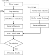
Alzheimer's disease is the neuro disorder which characterized by means of Amyloid- β (A β) in brain. However, accurate detection of this disease is a challenging task since the pathological issues of brain are complex in identification. In this paper, the changes associated with the retinal imaging for Alzheimer's disease are classified into two classes such as wild-type (WT) and transgenic mice model (TMM). For testing, optical coherence tomography (OCT) images are used to classify into two groups. The classification is implemented by support vector machines with the optimum kernel selection using a genetic algorithm. Among several kernel functions of SVM, the radial basis kernel function provides the better classification result. In order to deal with an effective classification using SVM, texture features of retinal images are extracted and selected. The overall accuracy reached 92% and 91% of precision for the classification of transgenic mice.
Copyright © 2022 Farrukh Sayeed et al.
Conflict of interest statement
The authors declare that there are no conflicts of interest.
Figures
Similar articles
-
Automated atrophy assessment for Alzheimer's disease diagnosis from brain MRI images.Magn Reson Imaging. 2019 Oct;62:167-173. doi: 10.1016/j.mri.2019.06.019. Epub 2019 Jul 4. Magn Reson Imaging. 2019. PMID: 31279772
-
Ensemble support vector machine classification of dementia using structural MRI and mini-mental state examination.J Neurosci Methods. 2018 May 15;302:66-74. doi: 10.1016/j.jneumeth.2018.01.003. Epub 2018 Feb 3. J Neurosci Methods. 2018. PMID: 29378218
-
Comparison of Feature Selection Techniques in Machine Learning for Anatomical Brain MRI in Dementia.Neuroinformatics. 2016 Jul;14(3):279-96. doi: 10.1007/s12021-015-9292-3. Neuroinformatics. 2016. PMID: 26803769
-
Frontiers for the Early Diagnosis of AD by Means of MRI Brain Imaging and Support Vector Machines.Curr Alzheimer Res. 2016;13(5):509-33. doi: 10.2174/1567205013666151116141705. Curr Alzheimer Res. 2016. PMID: 26567735 Review.
-
[Artificial Intelligence for Diagnostic Support of Neuroimage].Brain Nerve. 2019 Jul;71(7):733-748. doi: 10.11477/mf.1416201345. Brain Nerve. 2019. PMID: 31289247 Review. Japanese.
References
-
- Chibhabha F., Yang Y., Ying K., et al. Non-invasive optical imaging of retinal Aβ plaques using curcumin loaded polymeric micelles in APPswe/PS1ΔE9 transgenic mice for the diagnosis of Alzheimer’s disease. Journal of Materials Chemistry.B . 2020;8 - PubMed
MeSH terms
LinkOut - more resources
Full Text Sources
Medical


