

Case Reports
doi: 10.1016/j.radcr.2022.06.004.
eCollection 2022 Sep.
A rare case of recurrent primary dumbbell-shaped spinal hydatidosis
Affiliations
- PMID: 35814816
- PMCID: PMC9256548
- DOI: 10.1016/j.radcr.2022.06.004
Item in Clipboard
Case Reports
A rare case of recurrent primary dumbbell-shaped spinal hydatidosis
Radiol Case Rep.
.
Abstract
Spinal hydatidosis, which affects the thoracic vertebrae, is not only an extremely rare occurrence, but is also characterized by a high recurrence rate. Here, we reported a case of 67-years-old man who presented with recurrent spinal hydatid disease. The condition was originally misdiagnosed as Schwannoma via medical imaging, but eventually confirmed by postoperative pathology. He was subjected to surgery, combined with adjuvant drug therapy. Unfortunately, he experienced recurrent spinal hydatid disease and had to undergo hydatid cyst excision in over 5 years.
Keywords: Echinococcosis; Magnetic resonance imaging; Spine.
© 2022 The Authors.
Figures
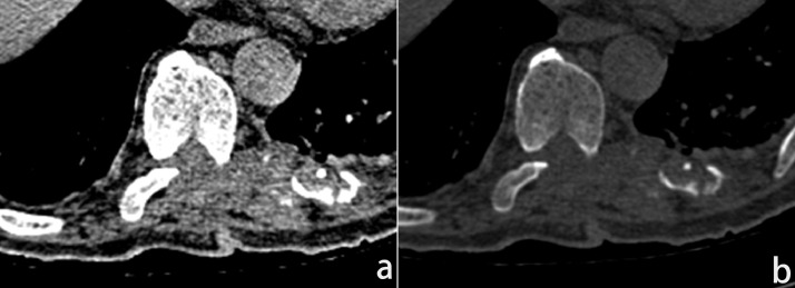
(a) CT soft tissue window. (b) CT bone window.
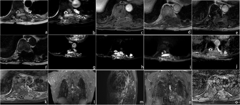
(a) MRI T2WI (axis image) showing hyperintense signal of a multilocular cystic cavity at the left side of the vertebral body. (b) MRI T2FS showing hyperintense of the cavity content. (c) TIWI Dixon sequence: WO image showing isointense signal of the lesion. (d) IP image showing hypointense. (e) OP image. (f) FO image showing hypointense. (g) MRI DWI (b = 0). (h) DWI (b = 600). (i) DWI (b = 1000). (g–i) showing hyperintense of the cavity. (j) MRI ADC map showing isointense. (k) Enhancement MRI (axis scan) showing the cystic cavity content without enhancement and the wall with slight enhancement. (l) Enhancement MRI (coronal scan) showing the cystic lesions in the spinal canal no enhancement. (m) Enhancement MRI (sagittal scan) showing the lesion ranges from T8 to T10 level. (n) Enhancement MRI (coronal scan, 2021) showing the lesion is larger, and the boundary with surrounding tissues unclear. (o) Enhancement MRI (axis scan, 2021) showing the cavity with more obvious enhancement.
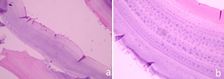
(a and b) Histopathological examination (Hematoxylin-Eosin staining): A thin membranous structure consisting of the outer and inner sacs can be seen (a) original magnification × 100. (b) Original magnification × 400).
Similar articles
-
Cauda equina syndrome caused by primary lumbosacral and pelvic hydatid cyst: a case report.Minim Invasive Neurosurg. 2007 Oct;50(5):292-5. doi: 10.1055/s-2007-973822. Minim Invasive Neurosurg. 2007. PMID: 18058646
-
Dumbbell hydatid cyst of the spine: case report and review of the literature.Spine (Phila Pa 1976). 2000 May 15;25(10):1296-9. doi: 10.1097/00007632-200005150-00018. Spine (Phila Pa 1976). 2000. PMID: 10806511 Review.
-
Disseminated intraspinal hydatid disease.J Neurosurg Spine. 2008 May;8(5):490-3. doi: 10.3171/SPI/2008/8/5/490. J Neurosurg Spine. 2008. PMID: 18447699
-
Case Report: Huge Dumbbell-Shaped Primary Hydatid Cyst Across the Intervertebral Foramen.Front Neurol. 2020 Nov 24;11:592316. doi: 10.3389/fneur.2020.592316. eCollection 2020. Front Neurol. 2020. PMID: 33329343 Free PMC article.
-
Recurrent spinal hydatidosis in North America. Case report and review of the literature.Neurosurg Focus. 2004 Dec 15;17(6):E8. doi: 10.3171/foc.2004.17.6.8. Neurosurg Focus. 2004. PMID: 15636578 Review.
References
-
- Turgut M. Hydatid disease of the spine: a survey study from Turkey. Infection. 1997;25(4):221–226. - PubMed
-
- Altinörs N, Bavbek M, Caner HH, Erdogan B. Central nervous system hydatidosis in Turkey: a cooperative study and literature survey analysis of 458 cases. J Neurosurg. 2000;93(1):1–8. - PubMed
-
- Pamir MN, Ozduman K, Elmaci I. Spinal hydatid disease. Spinal Cord. 2002;40(4):153–160. - PubMed
-
- Assefa G, Abebe M, Belete A, Schnider J. Epidural and para spinal thoracic hydatidosis presenting with progressive paraparesis and paraplegia: a case report. Ethiopian Med J. 2014;52(1):49–51. - PubMed
Publication types
LinkOut - more resources
Full Text Sources
