

Preliminary Cytomorphologic Diagnosis of Hematolymphoid Malignancies in Effusions: A Cyto-histo Correlation with Lessons on Restraint
- PMID: 35814877
- PMCID: PMC9262001
- DOI: 10.4103/joc.joc_204_21
Preliminary Cytomorphologic Diagnosis of Hematolymphoid Malignancies in Effusions: A Cyto-histo Correlation with Lessons on Restraint
Abstract
Background: Effusions as part of hematologic neoplasms are rare and as a primary presentation, rarer. In standalone laboratories of developing countries, resorting to techniques such as flow cytometry or immunohisto/cytochemistry may not be possible. A near definitive diagnosis on cytomorphology would, therefore, be an ideal beginning. To that end, we compiled our cases of primary hematolymphoid effusions, devising reproducible reporting categories and looked at their concordance with the final histopathology.
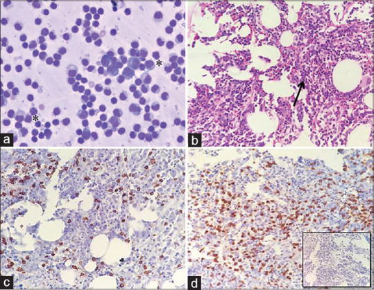
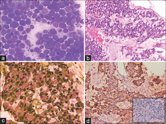
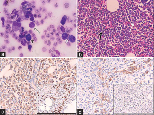
Subjects and methods: Fifty-four cases of primary hematolymphoid effusions over 10 years with cytology-histopathology correlation were chosen. Post morphology assessment, the cases were organized into six categories: suspicious of hematolymphoid malignancy, non-Hodgkin lymphoma-high-grade (NHL-HG), low-grade NHL (NHL-LG), Burkitt lymphoma, acute leukemias, and plasma cell dyscrasias. Discordance with histology was assigned as major and minor based mainly on therapeutic implications.
Results: Concordance was seen in a good number (81.5%) of cases. The NHL-HG and NHL-LG categories contributed to 33.3% each of major discordance. Tuberculosis and epithelial malignancies comprised the bulk of the major discordance. Overdiagnosis of a high-grade lymphoma for a histologically proven low-grade follicular lymphoma was the only case with minor discordance.
Conclusion: The cytologic categories used are not foolproof for hematologic neoplasms but have a fairly good concordance. A scanty abnormal population should always be viewed with suspicion and definitive labels should be avoided. While morphologic examination is fraught with danger, a good assessment directs the judicious selection of ancillary methods, and hence cannot be supplanted.
Keywords: Cytology; NHL; cytomorphology; effusion; hematologic neoplasms.
Copyright: © 2022 Journal of Cytology.
Conflict of interest statement
There are no conflicts of interest.
Figures
Similar articles
-
Lymphoreticular malignancies in serous effusions: Cytomorphologic, flow cytometric and immunocytochemical analysis.Diagn Cytopathol. 2021 May;49(5):647-656. doi: 10.1002/dc.24729. Epub 2021 Feb 25. Diagn Cytopathol. 2021. PMID: 33629825
-
Hematolymphoid neoplasms in effusion cytology.Cytojournal. 2018 Jun 14;15:15. doi: 10.4103/cytojournal.cytojournal_48_17. eCollection 2018. Cytojournal. 2018. PMID: 30034505 Free PMC article.
-
Combined cytomorphologic and immunophenotypic analysis in the diagnostic workup of lymphomatous effusions.Acta Cytol. 2001 May-Jun;45(3):307-12. doi: 10.1159/000327623. Acta Cytol. 2001. PMID: 11393059
-
Serous effusions in malignant lymphomas: a review.Diagn Cytopathol. 2006 May;34(5):335-47. doi: 10.1002/dc.20432. Diagn Cytopathol. 2006. PMID: 16604559 Review.
-
Value and limitations of fine-needle aspiration cytology in diagnosis and classification of lymphomas: A review.Diagn Cytopathol. 1999 Oct;21(4):240-9. doi: 10.1002/(sici)1097-0339(199910)21:4<240::aid-dc3>3.0.co;2-z. Diagn Cytopathol. 1999. PMID: 10495316 Review.
References
-
- Das DK, Al-Juwaiser A, George SS, Francis IM, Sathar SS, Sheikh ZA, et al. Cytomorphological and immunocytochemical study of non-Hodgkin's lymphoma in pleural effusion and ascitic fluid. Cytopathology. 2007;18:157–67. - PubMed
-
- Dermawan JKT, Policarpio-Nicolas ML. Malignancies in pleural, peritoneal, and pericardial effusions. Arch Pathol Lab Med. 2020;144:1086–91. - PubMed
-
- Gupta S, Sodhani P, Jain S. Cytomorphological profile of neoplastic effusions: An audit of 10 years with emphasis on uncommonly encountered malignancies. J Can Res Ther. 2012;8:602–9. - PubMed
-
- Naylor B. Pleural, Peritoneal and Pericardial effusions. In: Bibbo M, Wilbur DC, editors. Comprehensive Cytopathology. 3rd ed. Philadelphia, PA: Saunders/Elsevier; 2008. pp. 515–77.
LinkOut - more resources
Full Text Sources