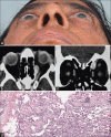
Intra-orbital fat necrosis following transcaruncular orbitotomy mimicking a surgical site infection: A case report
- PMID: 35814993
- PMCID: PMC9266475
- DOI: 10.4103/1319-4534.347307
Intra-orbital fat necrosis following transcaruncular orbitotomy mimicking a surgical site infection: A case report
Abstract
Fat necrosis is a benign non-suppurative inflammation of adipose tissue most commonly occurring in breast, subcutaneous tissue or intraabdominal fat post trauma, surgery, radiation. Transcaruncularorbitotomy provides a safe, rapid, and cosmetically pleasing approach to the medial wall and orbital apex. Intraorbital fat necrosis as its complication has not been documented in literature. The authors report the case of an elderly lady who presented with localized pain, swelling following a transcaruncular orbitotomy for excision biopsy of an orbital vascular mass. The etiology, clinical presentation, intraoperative finding, imaging, and possible mechanisms contributing to the pathogenesis of postsurgical orbital fat necrosis has been suggested.
Keywords: Orbit; Transcaruncular orbitotomy; fat necrosis; non encapsulated.
Copyright: © 2022 Saudi Journal of Ophthalmology.
Conflict of interest statement
There are no conflicts of interest.
Figures


Similar articles
-
The transcaruncular approach to the medial orbital wall.Laryngoscope. 2002 Jun;112(6):986-9. doi: 10.1097/00005537-200206000-00009. Laryngoscope. 2002. PMID: 12160296
-
[Transcaruncular medial orbitotomy--a case report].Cesk Slov Oftalmol. 2011 Nov-Dec;67(5-6):178-80. Cesk Slov Oftalmol. 2011. PMID: 22448419 Slovak.
-
Transcaruncular approach to the medial orbit and orbital apex.Ophthalmology. 2000 Aug;107(8):1459-63. doi: 10.1016/s0161-6420(00)00241-4. Ophthalmology. 2000. PMID: 10919889
-
[Excisional surgery of orbital tumors].Ophthalmologe. 2021 Oct;118(10):995-1003. doi: 10.1007/s00347-021-01386-5. Epub 2021 Apr 23. Ophthalmologe. 2021. PMID: 33893529 Review. German.
-
Visceral adiposity and inflammatory bowel disease.Int J Colorectal Dis. 2021 Nov;36(11):2305-2319. doi: 10.1007/s00384-021-03968-w. Epub 2021 Jun 9. Int J Colorectal Dis. 2021. PMID: 34104989 Review.
Cited by
-
Spectrum of orbital fat necrosis in rhino-orbital-cerebral mucormycosis in post-COVID-19 patients.Indian J Ophthalmol. 2024 Oct 1;72(10):1478-1482. doi: 10.4103/IJO.IJO_2623_23. Epub 2024 Sep 27. Indian J Ophthalmol. 2024. PMID: 39331438 Free PMC article.
References
-
- Tan PH, Lai LM, Carrington EV, Opaluwa AS, Ravikumar KH, Chetty N, et al. Fat necrosis of the breast–A review. Breast. 2006;15:313–8. - PubMed
-
- Chala LF, de Barros N, de Camargo Moraes P, Endo E, Kim SJ, Pincerato KM, et al. Fat necrosis of the breast: Mammographic, sonographic, computed tomography, and magnetic resonance imaging findings. Curr Probl Diagn Radiol. 2004;33:106–26. - PubMed
-
- Takami H, Asai A, Ohshige H, Uesaka T, Takeda J, Yoshimura K, et al. Fat necrosis appearing as intraorbital tumour: Case report. Br J Neurosurg. 2014;28:525–7. - PubMed
-
- Graham SM, Thomas RD, Carter KD, Nerad JA. The transcaruncular approach to the medial orbital wall. Laryngoscope. 2002;112:986–9. - PubMed
-
- Caucanas M, Heureux F, Müller G, Vanhooteghem O. Atypical hypodermic necrosis secondary to insulin injection: A case report and review of the literature. J Diabetes. 2011;3:19–20. - PubMed
Publication types
LinkOut - more resources
Full Text Sources
Miscellaneous
