. 2022 May 14;7(7):265-267.
doi: 10.1016/j.vgie.2022.03.010. eCollection 2022 Jul.

Purely endoscopic appendectomy

Affiliations

Purely endoscopic appendectomy

Sergey V Kantsevoy et al. VideoGIE. .

Abstract

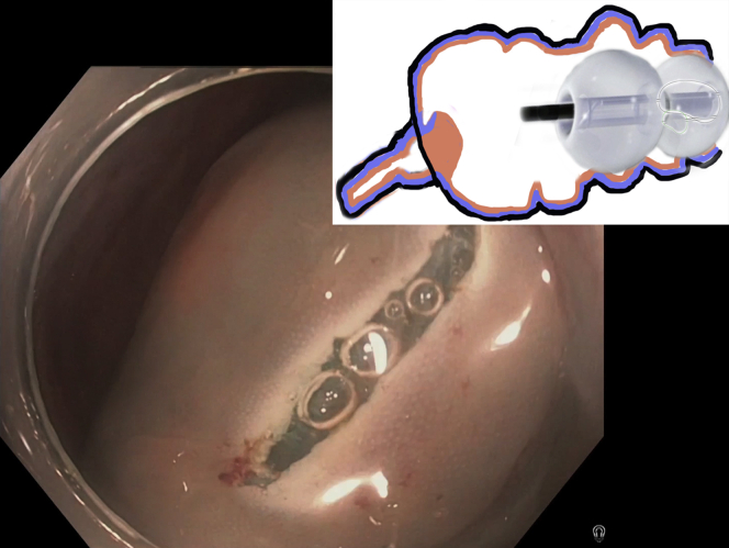
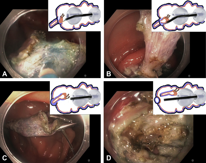
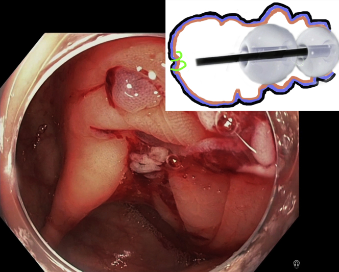
Video 1Submucosal nodule in the cecum. After submucosal injection, a circumferential incision of the mucosa surrounding the lesion is performed with DualKnife (Olympus America, Center Valley, Pa, USA). The fore-balloon of the double-balloon endoluminal interventional platform (DBEIP) is deployed and the edges of the circumferential incision are attached with 2 endoscopic clips to the long suture-loop mounted on the fore-balloon of the DBEIP. The fore-balloon is retracted in anal direction, pulling the lesion into the cecum. Careful endoscopic submucosal dissection is performed with DualKnife and HookKnife (Olympus America). Dissection is markedly facilitated by traction and continued until the entire appendix is pulled into the cecum. The tip of the appendix is separated from surrounding tissues, resulting in a full-thickness cecal wall defect. The suture-loop holding the resected appendix is cut with LoopCutter (Olympus America). The resected appendix is removed through DBEIP and the Overstitch endoscopic suturing device (Apollo Endosurgery, Austin, Tex, USA) is advanced into the cecum. The full-thickness defect in the cecal wall is completely closed with 2 continuous sutures. The final view demonstrates the entire resected appendix.

Keywords: DBEIP, double-balloon endoluminal interventional platform.


Figures

Figure 1
Figure 1
Cecal submucosal lesion. In the accompanying diagram, the mucosal layer is orange, submucosal layer is blue, and serosal layer is black.
Figure 2
Figure 2
Circumferential incision around the lesion is performed with DualKnife (Olympus America, Center Valley, Pa, USA). In the accompanying diagram, the mucosal layer is orange, submucosal layer is blue, and serosal layer is black.
Figure 3
Figure 3
The lesion is attached with endoscopic clips (Resolution 360, Boston Scientific Corporation, Natick, Mass, USA) to a suture-loop mounted on fore-balloon of the double-balloon endoluminal interventional platform and pulled into the colonic lumen. In the accompanying diagram, the mucosal layer is orange, submucosal layer is blue, and serosal layer is black.
Figure 4
Figure 4
The lesion is carefully dissected from surrounding tissues. In the accompanying diagrams, the mucosal layer is orange, submucosal layer is blue, and serosal layer is black. A, Endoscopic submucosal dissection is started with Dual Knife. B, The dissection continued with HookKnife (Olympus America, Center Valley, Pa, USA). C, The entire appendix is pulled into the cecum and dissected from surrounding tissues. D, Full-thickness defect in cecal wall after dissection is completed and the appendix is separated from the cecum.
Figure 5
Figure 5
The entire appendix is resected and removed en bloc.
Figure 6
Figure 6
Overstitch endoscopic suturing device (Apollo Endosurgery, Austin, Tex, USA) is delivered to the cecum through the double-balloon endoluminal interventional platform. In the accompanying diagram, the mucosal layer is orange, submucosal layer is blue, and serosal layer is black.
Figure 7
Figure 7
Full-thickness defect post appendectomy is completely closed with 2 continuous sutures. In the accompanying diagram, the mucosal layer is orange, submucosal layer is blue, and serosal layer is black.

References

    1. Dotai T., Coker A.M., Antozzi L., et al. Transgastric large-organ extraction: the initial human experience. Surg Endosc. 2013;27:394–399. - PubMed
    1. Auyang E.D., Santos B.F., Enter D.H., et al. Natural orifice translumenal endoscopic surgery (NOTES®): a technical review. Surg Endosc. 2011;25:3135–3148. - PubMed
    1. Coomber R.S., Sodergren M.H., Clark J., et al. Natural orifice translumenal endoscopic surgery applications in clinical practice. World J Gastrointest Endosc. 2012;4:65–74. - PMC - PubMed
    1. Huang C., Huang R.X., Qiu Z.J. Natural orifice transluminal endoscopic surgery: new minimally invasive surgery come of age. World J Gastroenterol. 2011;17:4382–4388. - PMC - PubMed
    1. Palanivelu C., Rajan P.S., Rangarajan M., et al. Transvaginal endoscopic appendectomy in humans: a unique approach to NOTES-world's first report. Surg Endosc. 2008;22:1343–1347. - PubMed

LinkOut - more resources