Review
. 2022 Jun 22:10:879609.
doi: 10.3389/fchem.2022.879609. eCollection 2022.

Rhodopsins: An Excitingly Versatile Protein Species for Research, Development and Creative Engineering

Affiliations
Review

Rhodopsins: An Excitingly Versatile Protein Species for Research, Development and Creative Engineering

Willem J de Grip et al. Front Chem. .

Abstract

The first member and eponym of the rhodopsin family was identified in the 1930s as the visual pigment of the rod photoreceptor cell in the animal retina. It was found to be a membrane protein, owing its photosensitivity to the presence of a covalently bound chromophoric group. This group, derived from vitamin A, was appropriately dubbed retinal. In the 1970s a microbial counterpart of this species was discovered in an archaeon, being a membrane protein also harbouring retinal as a chromophore, and named bacteriorhodopsin. Since their discovery a photogenic panorama unfolded, where up to date new members and subspecies with a variety of light-driven functionality have been added to this family. The animal branch, meanwhile categorized as type-2 rhodopsins, turned out to form a large subclass in the superfamily of G protein-coupled receptors and are essential to multiple elements of light-dependent animal sensory physiology. The microbial branch, the type-1 rhodopsins, largely function as light-driven ion pumps or channels, but also contain sensory-active and enzyme-sustaining subspecies. In this review we will follow the development of this exciting membrane protein panorama in a representative number of highlights and will present a prospect of their extraordinary future potential.

Keywords: eukaryotic; ion pumps; membrane protein; microbial; optogenetics; photoreceptor; retinal protein; visual pigments.


Conflict of interest statement

The authors declare that the research was conducted in the absence of any commercial or financial relationships that could be construed as a potential conflict of interest.

Figures

FIGURE 1
FIGURE 1
Chemical structures of the most common chromophore configurations in the rhodopsin families. The type-2 pigments contain an 11-cis, 15-anti retinylidene Schiff base of retinal A1 (A) in the “dark state” (or “ground state” in photophysical terminology), which is photo-excited into the all-trans configuration. The Type-1 pigments contain the all-trans configuration (B) in the “dark state.” This is photo-excited into the 13-cis, 15-anti configuration (C), which thermally relaxes and re-isomerizes, returning to the ground state. The ring-polyene chain orientation is different for type-2 (6-s-cis) and type-1 (6-s-trans) rhodopsins.
FIGURE 2
FIGURE 2
Schematic of a vertebrate rod photoreceptor cell (scotopic vision), zooming in on the location of the rod visual pigment rhodopsin. The rod outer segment (ROS), a ciliary outgrowth, is densely filled with isolated flattened vesicles (discs) which contain rhodopsin as the major (ca 90% w/w) membrane protein. The vertebrate visual pigments are therefore also designated as “ciliary rhodopsins.” Other disc membrane proteins are involved in signal propagation, stabilization of the disc shape and communication with the plasma membrane (PM). The phospholipids in the disc membrane have an exceptionally high content (ca 40%) of highly unsaturated fatty acids (22:6∞3) (Daemen, 1973). The discs are continuously generated at the base of the ROS as invaginations of the PM, then are nipped off and move upwards. After 7–10 days they reach the top of the ROS, which is pinched off in a circadian rhythm and degraded in the adjacent retinal pigment epithelium (RPE) (Young, 1976). The vertebrate cone photoreceptor (photopic vision) is organized in a similar fashion, except that the “discs” remain continuous with the PM as invaginations and are not pinched off. The organization of invertebrate visual photoreceptors is roughly similar, but the photoreceptive membranes are organized as numerous microvilli in rhabdomeric structures (Warrant and McIintyre, 1993) and their rhodopsins are also designated as rhabdomeric visual pigments. Only the classical visual pigments (Opn1, Opn2 and R-gene families) are organized in these specialized cellular outgrowths. All other type-2 and all type-1 pigments are targeted to the PM or an eyespot and form only a small part (up to several percent) of that membrane protein population.
FIGURE 3
FIGURE 3
Typical dark state absorbance spectra (red curves) of a purified type-2 (A) and type-1 (B) pigment. Both spectra exhibit a major peak (α-band) and a small satellite (β-band), both originating in the chromophore, and a γ-band near 280 nm, mainly originating in protein residues. The α-band derives from the whole conjugated polyene system (S0-S1) (cf. Figure 1), while the β-band derives from a smaller segment, and its intensity also depends on the torsion in the polyene chain. Upon short illumination of the monostable type-2 pigment (A) in the presence of hydroxylamine, the liberated retinal is converted into retinaloxime (blue curve). The Meta state of bistable type-2 pigments, also reacts with hydroxylamine generating retinaloxime, but usually quite slowly. Short illumination of type-1 pigments (B) in the presence of hydroxylamine hardly affects the photocycle and the return to the ground state. However, upon prolonged illumination hydroxylamine will slowly attack photo-intermediates, mainly M and N, releasing retinaloxime.
FIGURE 4
FIGURE 4
Membrane mimics for purified membrane proteins. Schematics of the micellar (A), nanodisc (B) and vesicular (C; proteoliposome) organization are displayed. Note that the relative dimensions are not to scale: diameters vary from 10–50 nm for the micelles and nanodiscs, and from 100 nm up to 10 µm for the liposomes. Several amphipatic components functioning as bilayer-stabilizing agents in the nanodiscs have been generated (MSP derivatives from lipoproteins, synthetic amphipols and SMAs, respectively), and are still under further development. Purification of the protein in a detergent environment generates the classical micellar state (A). Because the thermal stability of membrane proteins in the micelles is generally reduced, often (phospho)lipids are added (bicelles). Alternatively, membrane proteins can be transferred into the bilayer membrane of a nanodisc (B) or liposome (C). Membrane proteins can be directly (amphipol or SMA nanodiscs) or under very brief detergent exposure (MSP nanodiscs) transferred from the native membrane into nanodiscs, and the classical purification techniques can be applied upon the resulting nanodisc population. Liposomes offer a broader selection for the lipid population, and are used in vectorial transport studies and in AFM, FTIR and solid-state NMR spectroscopy. However, they are less suitable in optical spectroscopy because of their larger dimension, resulting in strong light scattering.
FIGURE 5
FIGURE 5
Some uncommon retinal analogs occurring as natural chromophores or in engineered pigment analogs. 3, 4-didehydroretinal (retinal A2, (A)) red-shifts the rhodopsin spectrum relative to A1, and is mostly found in fish and amphibian visual pigments. 3-hydroxy- (B) and 4-hydroxy- (D) retinal A1 induce a blue-shift relative to A1 and are found in the visual pigments of insects and deep-sea shrimps, respectively. Phenylretinal (F), MMAR (C) and the merocyanine derivative (E) are synthetic analogs, that, respectively, induce a blue-shift (F) and the largest red-shifts, observed so far ((C,E); see text). All these analogs bind to the lysine residue in the native opsin binding pocket with a protonated Schiff base.
FIGURE 6
FIGURE 6
Comparison of structural features of the type-2 and type-1 pigment archetypes bovine rod rhodopsin (left section) and bacteriorhodopsin (right section), respectively. Full crystal structures are presented in (A) and (A) (pdb 1U19 and 5ZIN), a top view is shown in (B) and (B) and a binding pocket exposure in (C) and (C), respectively. The retinylidene chromophore (cyan) is represented as space-filling spheres, and the retinal binding lysine residue (cyan) is presented as sticks. The two protein residues displayed (red) contribute to the counterion complex stabilizing the pronated Schiff base. The two crystal structures share the seven α-helical transmembrane segment bundle, but the packing of the helices, the location and assembly of the binding pocket and the structure of the chromophore are clearly different.
FIGURE 7
FIGURE 7
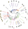
Global phylogeny of type-1 pigments illustrating their formidable diversification. The figure was modified with consent from Rozenberg et al., 2021. We refer to the original paper for the construction of the tree and for all abbreviations. Purple arrows represent active outward (away from center) and inward ion transport, respectively. Orange arrows represent ion channels. Pink arrows represent enzyme-rhodopsins (fused enzyme domains) and sensory rhodopsins (detachable transducers). For further details of the various classes we refer to recent literature (Hasemi et al., 2016; Govorunova et al., 2016; id-, 2022; Nakajima et al., 2018; Oppermann et al., 2019; Kovalev et al., 2020a; Rozenberg et al., 2021).
FIGURE 8
FIGURE 8
Global presentation of the predominant photochemical pathways in the rhodopsin families. (A) Bovine rod rhodopsin as the archetype of the monostable type-2 pigments, (B) squid/fly visual pigment chimera, typical for the bistable type-2 pigments, and (C) bacteriorhodopsin (BR) as a prototype for the type-1 pigments. The “dark state” 11-cis, 15-anti chromophore configuration in type-2 pigments is photo-excited into all-trans. The all-trans chromophore configuration in type-1 pigments is photo-excited into 13-cis, 15-anti, which thermally relaxes, eventually returning to the ground state. The early photo-intermediates still contain a protonated Schiff base and relax thermally to deprotonated Meta II or M states. In proton pumps like BR this is accompanied by opening up proton pathways in the protein, while in most type-2 pigments binding of a G protein is initiated. At this stage, the pathways divert, as further explained in the text. Of course, here are exceptions: some type-2 pigments contain all-trans in the “dark state,” which is photoexcited into 11-cis, either to change activity or to release 11-cis retinal for regeneration of visual opsins (Table 2). Some type-1 pigments can also photo-generate 9-cis or 11-cis states with deviating photocycles and/or functions. (D) Simplified schematic of a conical intersection where the excited chromophore at the S1 energy surface can cross over to the S0 energy surface of the photoproduct. The S1 surface can contain thermal transitions, and in type-1 pigments the kinetics to reach and cross-over at the conical intersection also depend on the pKa of the direct counterion to the Schiff base (Chang et al., 2022).

Similar articles

Cited by

References

    1. Abdelfattah A. S., Farhi S. L., Zhao Y., Brinks D., Zou P., Ruangkittisakul A., et al. (2016). A Bright and Fast Red Fluorescent Protein Voltage Indicator that Reports Neuronal Activity in Organotypic Brain Slices. J. Neurosci. 36, 2458–2472. 10.1523/jneurosci.3484-15.2016 - DOI - PMC - PubMed
    1. Abdelfattah A. S., Valenti R., Zheng J., Wong A., Chuong A. S., Hasseman J. P., et al. (2020). A General Approach to Engineer Positive-Going eFRET Voltage Indicators. Nat. Commun. 11, 3444–34413448. 10.1038/s41467-020-17322-1 - DOI - PMC - PubMed
    1. Abdulaev N. G., Artamonov I. D., Bogachuk A. S., Feigina M. Y., Kostina M. B., Kudelin A. B., et al. (1982). Structure of Light-Activated Proteins - Visual Rhodopsin. Biochem. Int. 5, 693–703.
    1. Abdulaev N. G., Ridge K. D. (2000). Heterologous Expression of Bovine Opsin in Pichia pastoris . Meth. Enzymol. 315, 3–11. 10.1016/s0076-6879(00)15831-8 - DOI - PubMed
    1. Acharya A. R., Vandekerckhove B., Larsen L. E., Delbeke J., Wadman W. J., Vonck K., et al. (2021). In Vivo blue Light Illumination for Optogenetic Inhibition: Effect on Local Temperature and Excitability of the Rat hippocampus. J. Neural Eng. 18, 066038–066031. 10.1088/1741-2552/ac3ef4 - DOI - PubMed

LinkOut - more resources