. 2023 Mar 31;119(2):506-519.
doi: 10.1093/cvr/cvac108.

Nogo-A reduces ceramide de novo biosynthesis to protect from heart failure

Affiliations

Nogo-A reduces ceramide de novo biosynthesis to protect from heart failure

Linda Sasset et al. Cardiovasc Res. .

Abstract

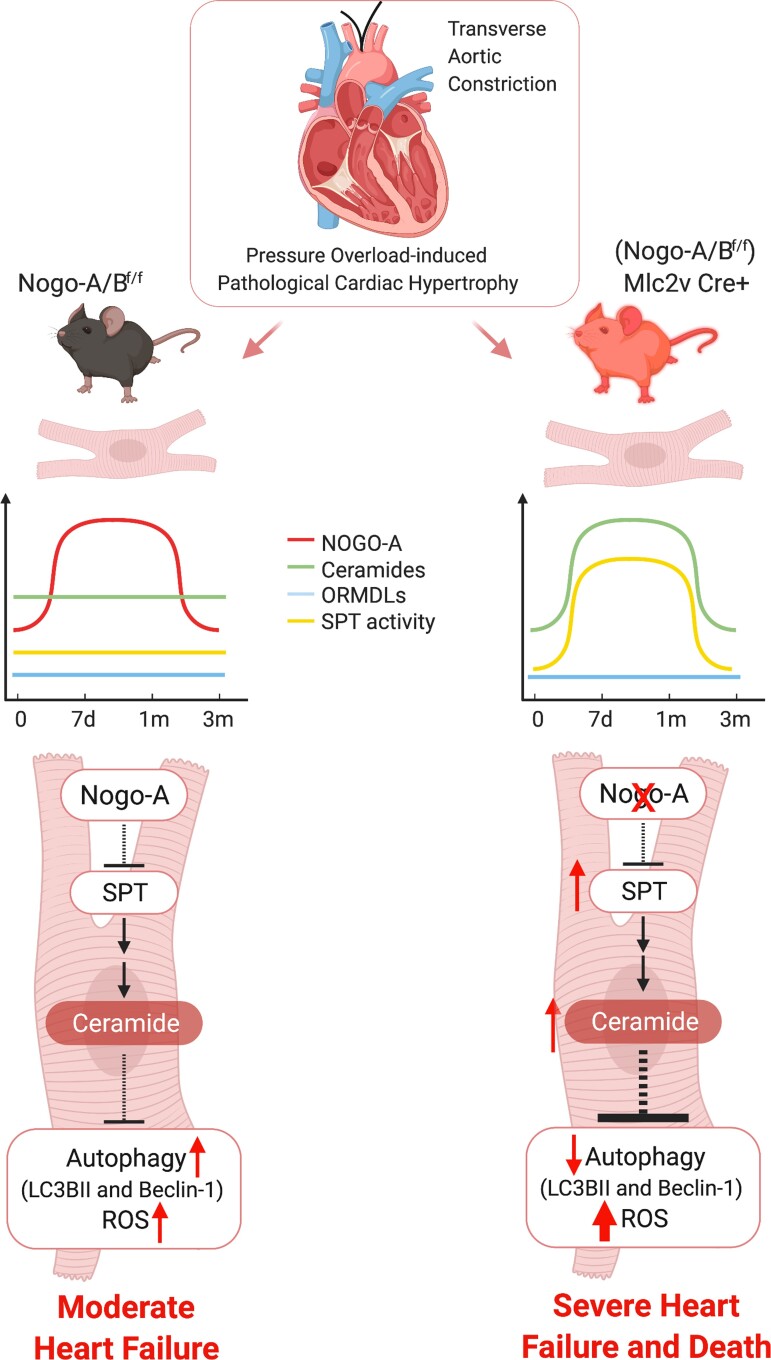
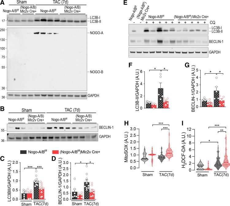
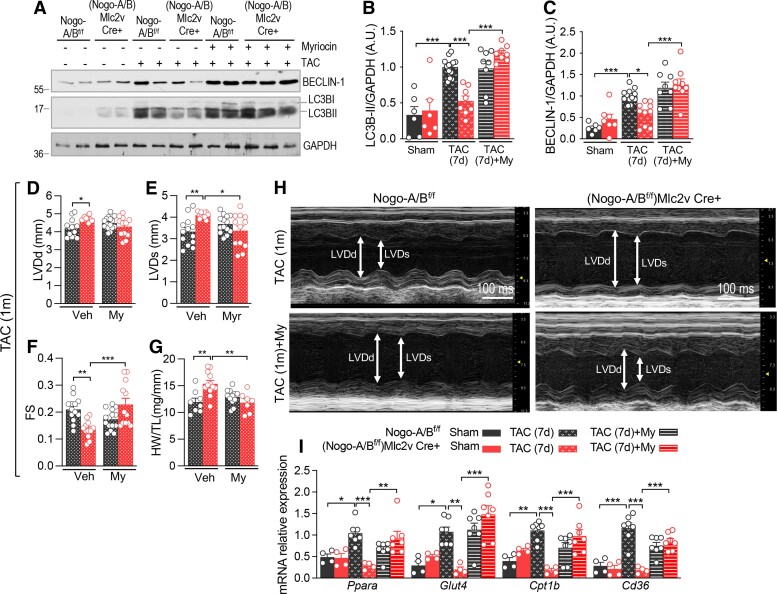
Aims: Growing evidence correlate the accrual of the sphingolipid ceramide in plasma and cardiac tissue with heart failure (HF). Regulation of sphingolipid metabolism in the heart and the pathological impact of its derangement remain poorly understood. Recently, we discovered that Nogo-B, a membrane protein of endoplasmic reticulum, abundant in the vascular wall, down-regulates the sphingolipid de novo biosynthesis via serine palmitoyltransferase (SPT), first and rate liming enzyme, to impact vascular functions and blood pressure. Nogo-A, a splice isoform of Nogo, is transiently expressed in cardiomyocyte (CM) following pressure overload. Cardiac Nogo is up-regulated in dilated and ischaemic cardiomyopathies in animals and humans. However, its biological function in the heart remains unknown.

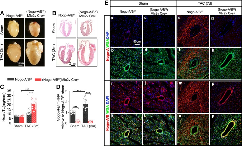
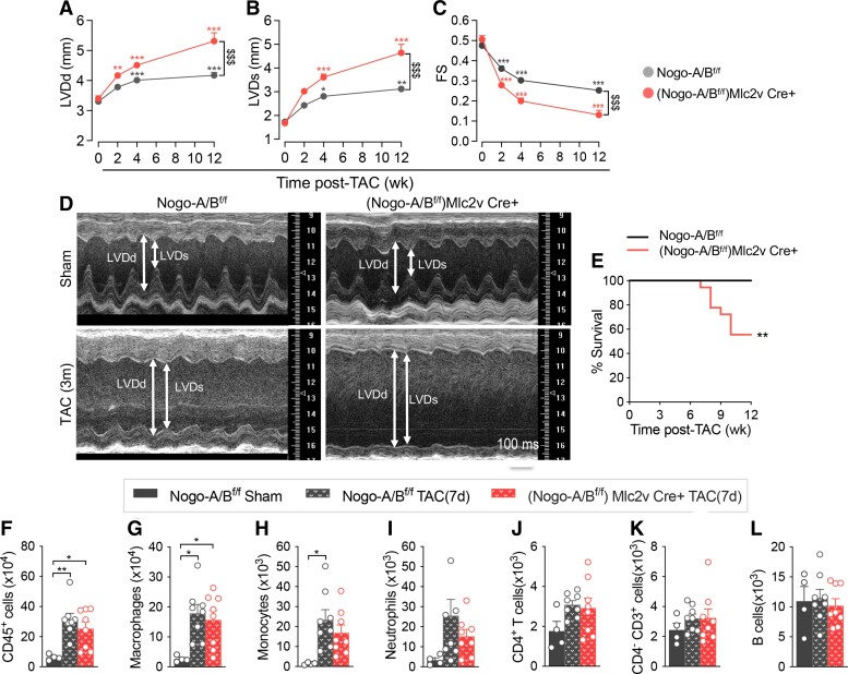
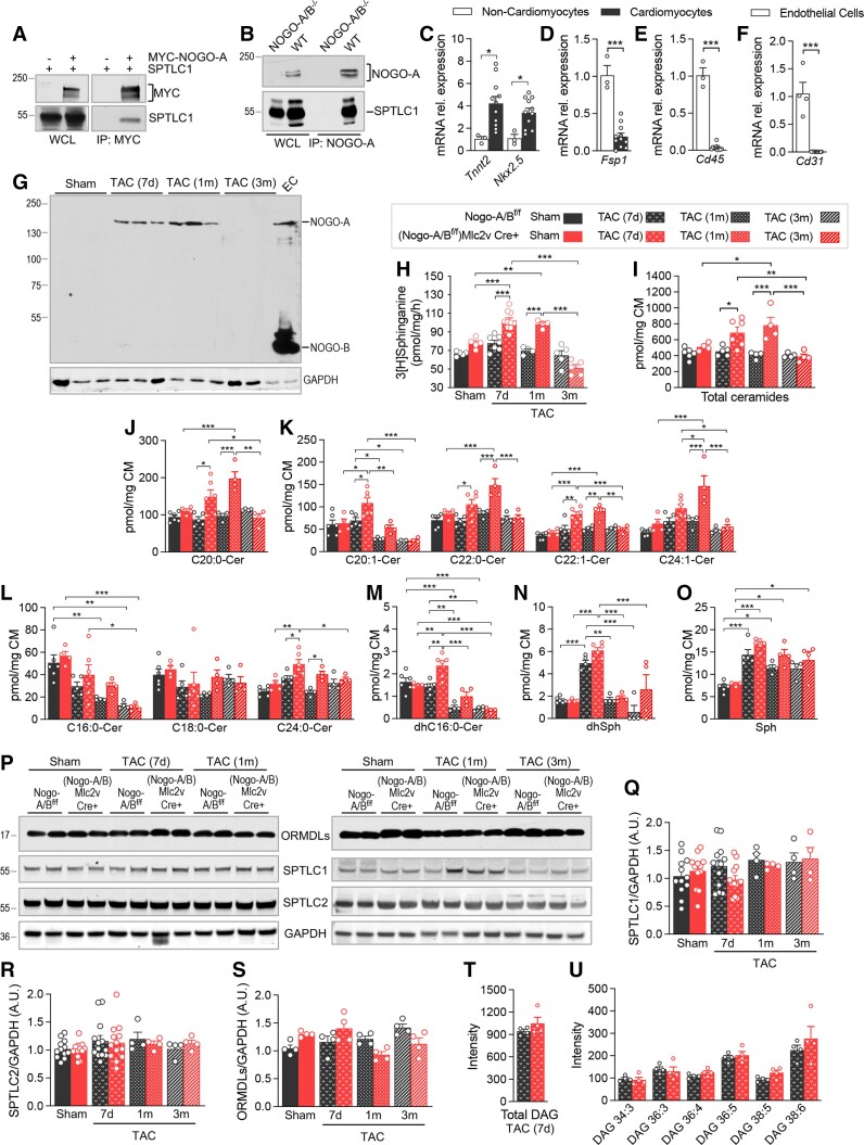
Methods and results: We discovered that Nogo-A is a negative regulator of SPT activity and refrains ceramide de novo biosynthesis in CM exposed to haemodynamic stress, hence limiting ceramide accrual. At 7 days following transverse aortic constriction (TAC), SPT activity was significantly up-regulated in CM lacking Nogo-A and correlated with ceramide accrual, particularly very long-chain ceramides, which are the most abundant in CM, resulting in the suppression of 'beneficial' autophagy. At 3 months post-TAC, mice lacking Nogo-A in CM showed worse pathological cardiac hypertrophy and dysfunction, with ca. 50% mortality rate.

Conclusion: Mechanistically, Nogo-A refrains ceramides from accrual, therefore preserves the 'beneficial' autophagy, mitochondrial function, and metabolic gene expression, limiting the progression to HF under sustained stress.

Keywords: Autophagy; Ceramide; Heart failure; Mitochondrial function; Sphingolipid metabolism.


Conflict of interest statement

Conflict of interest: None declared.

Figures

Graphical abstract
Graphical abstract
Figure 1
Figure 1
The loss of cardiomyocytes Nogo-A exacerbates pathological cardiac hypertrophy. Representative images of (A) whole heart and (B) H&E staining of longitudinal heart sections of Nogo-A/Bf/f and (Nogo-A/Bf/f)Mlc2v-Cre+ sham and 3-month TAC-operated mice. (C) Heart weight/tibial length (HW/TL) ratios of sham and 3-month TAC-operated mice. (D) Real-time (RT)-PCR for Nogo-A/B mRNA expression in CM isolated from sham and 7 days TAC-operated Nogo-A/Bf/f and (Nogo-A/Bf/f)Mlc2v-Cre+ mice. GAPDH was used as housekeeping gene (n = 7 mice/group). (E) Immunofluorescence staining for Nogo-A/B and Nogo-A of myocardial sections from sham and TAC-operated Nogo-A/Bf/f and (Nogo-A/Bf/f)Mlc2v-Cre+ mice. FITC-labelled wheat germ agglutinin (WGA) staining has been used to evidence cell membranes in myocardial sections. Nuclei are stained with DAPI. Data are expressed as mean ± SEM. ***P≤0.001; Statistical significance was determined by two-way ANOVA followed by Tukey’s multiple comparison test.
Figure 2
Figure 2
The absence of Nogo-A in CM worsen heart failure and decreases survival rate. Echocardiographic analysis of sham- and TAC-operated Nogo-A/Bf/f (n = 14) and (Nogo-A/Bf/f)Mlc2v-Cre+ (n = 12) mice at different time points. (A) Left-ventricle end-diastolic diameter (LVDd), (B) LV end-systolic diameter (LVDs), (C) fractional shortening (FS) measured at baseline and 2, 4, and 12 weeks after TAC. (D) Representative images of LV serial echocardiography. (E) Survival curve of Nogo-A/Bf/f (n = 16) and (Nogo-A/Bf/f)Mlc2v-Cre+ (n = 18) post-TAC. Survival analysis was performed by the Kaplan–Meier method, and between-group differences in survival were evaluated by using the Gehan–Breslow–Wilcoxon test. Flow cytometry analysis of inflammatory cells in non-CM cell suspension from the hearts of sham-operated Nogo-A/Bf/f mice (n = 4), Nogo-A/Bf/f and (Nogo-A/Bf/f)Mlc2v-Cre+ at 7 days post-TAC (n = 8 mice/group). Total number of: (F) CD45+ haematopoietic cells; (G) CD45+, CD64Lo, Ly6CHi monocytes; (H) CD45+, Ly6CLo, Ly6G-, CD64Hi macrophages; (I) CD45+, CD64-, Ly6CMed, Ly6GHi neutrophils; (J) CD4+ CD3+ T cells; (K) CD4- CD3+ T cells; and (L) CD19+ B cells. Data are expressed as mean ± SEM. *P≤0.05; **P≤0.01. In A–C, *** is statistic vs. time 0 of the same genotype, and $$$ is statistic between the genotypes. Statistical significance was determined by two-way ANOVA followed by Tukey’s multiple comparison test (A–C) and one-way ANOVA (F–L)
Figure 3
Figure 3
Nogo-A interacts with SPT and regulates ceramide de novo biosynthesis in cardiomyocytes. (A) Western blot (WB) analysis of transfected SPTLC1 and MYC-NOGO-A in HEK293T whole cell lysate (WCL) and after co-immunoprecipitation (co-IP) with anti-MYC antibody. (B) WB analysis of Nogo-A and SPTLC1 in NOGO-A/B-/- and WT myocardial microsomes, in WCL and after co-IP with anti-NOGO-A antibody. RT-PCR for (C) Tnnt2 and Nkx2.5, (D) Fsp1, and (E) Cd45 mRNA expression in CM vs. non-CM fractions. (F) RT-PCR for Cd31 mRNA expression in CM fraction vs. endothelial cells (ECs). (G) WB analysis of NOGO-A in CM isolated from Nogo-A/Bf/f and (Nogo-A/Bf/f)Mlc2v-Cre+ mice sham, 7 days, 1 and 3 months post-TAC. EC lysate has been used as positive control for NOGO-B expression. (H) SPT activity in CM isolated from Nogo-A/Bf/f [sham n = 5; TAC (7d) n = 7; TAC (1m) n = 4; TAC (3m) n = 5] and (Nogo-A/Bf/f)Mlc2v-Cre+ [sham n = 5; TAC (7d) n = 10; TAC (1m) n = 4; TAC (3m) n = 5] mice. (I) Total ceramides, (J–L) specific ceramides, (M) C16:0-dihydro ceramide (dhC16:0-cer), (N) dihydrosphingosine (dhSph), and (O) sphingosine (Sph) measured in CM isolated from Nogo-A/Bf/f [sham n = 6; TAC (7d) n = 5, TAC (1m) n = 4, TAC (3m) n = 4] and (Nogo-A/Bf/f)Mlc2v-Cre+ [sham n = 4; TAC (7d) n = 6, TAC (1m) n = 4, TAC (3m) n = 4] mice. (P) WB analysis and (Q–S) relative quantification of ORMDLs, SPTLC1, and SPTLC2 expression, performed on CM isolated from sham and TAC(7d)-operated Nogo-A/Bf/f and (Nogo-A/Bf/f)Mlc2v-Cre+ mice (n ≥ 4/group). GAPDH was used as housekeeping. (T) Total diacylglycerols (DAGs) and (U) specific DAG measured in Nogo-A/Bf/f and (Nogo-A/Bf/f)Mlc2v-Cre+ (n = 4/group) CM at 7 days post-TAC. Data are expressed as mean ± SEM. *P≤0.05; **P≤0.01; ***P≤0.001. Statistical significance was determined by unpaired t test (C–F, T–U), and two-way ANOVA followed by Tukey’s multiple comparison test (H–O, Q–S).
Figure 4
Figure 4
Cardiomyocyte Nogo-A preserves beneficial autophagy by refraining ceramide de novo biosynthesis. (A) WB analysis of LC3B-II and NOGO-A/B and (B) BECLIN-1 expression in CM from Nogo-A/Bf/f and (Nogo-A/Bf/f)Mlc2v-Cre+ mice. GAPDH was used as housekeeping. (C) Quantification of LC3B-II expression in Nogo-A/Bf/f [sham n = 7; TAC (7d) n = 11] and (Nogo-A/Bf/f)Mlc2v-Cre+ [sham n = 7; TAC (7d) n = 12] CM. (D) Quantification of BECLIN-1 expression in Nogo-A/Bf/f [sham n = 6; TAC (7d) n = 10] and (Nogo-A/Bf/f)Mlc2v-Cre+ [sham n = 6; TAC (7d) n = 8] CM. (E) WB analysis of LC3B-II and BECLIN-1 expression in CM from Nogo-A/Bf/f and (Nogo-A/Bf/f)Mlc2v-Cre+ mice at 7 days post-TAC, with or without CQ. GAPDH was used as housekeeping. Quantification of (F) LC3B-II and (G) BECLIN-1 expression in Nogo-A/Bf/f and (Nogo-A/Bf/f)Mlc2v-Cre+ CM (n = 5). Quantification of (H) mitochondrial ROS in Nogo-A/Bf/f [sham n = 16; TAC (7d) n = 91] and (Nogo-A/Bf/f)Mlc2v-Cre+ [sham n = 109; TAC (7d) n = 59] CM and (I) cellular ROS in Nogo-A/Bf/f [sham n = 11; TAC (7d) n = 153] and (Nogo-A/Bf/f)Mlc2v-Cre+ [sham n = 77; TAC (7d) n = 86] CM. Data are expressed as mean ± SEM. *P≤0.05; **P≤0.01; ***P≤0.001. Statistical significance was determined by two-way ANOVA followed by Tukey’s multiple comparison test.
Figure 5
Figure 5
Inhibition of the ceramide de novo biosynthesis with myriocin restores CM autophagy and prevents HF in absence of Nogo-A. (A) WB analysis and quantification of (B) LC3B-II and (C) BECLIN-1 expression in CM from Nogo-A/Bf/f [sham n = 6, TAC (7d) n = 12, TAC (7d + My) n = 8] and (Nogo-A/Bf/f)Mlc2v-Cre+ [sham n = 6, TAC (7d) n = 8, TAC (7d + My) n = 8] mice. GAPDH was used as housekeeping. Echocardiographic analysis of vehicle- and My-treated TAC-operated Nogo-A/Bf/f (vehicle n = 13; My n = 15) and (Nogo-A/Bf/f)Mlc2v-Cre+ (vehicle n = 12; My n = 12) mice. (D) LVDd, (E) LVDs, (F) FS, and (G) HW/TL ratios of vehicle- and My-treated Nogo-A/Bf/f (vehicle n = 8; My n = 12) and (Nogo-A/Bf/f)Mlc2v-Cre+ mice (vehicle n = 12, My n = 7) measured at 1 month post-TAC. (H) Representative images of LV serial echocardiography. (I) RT-PCR for Ppara, Glut4, Cpt1B, and Cd36 in CM isolated from Nogo-A/Bf/f [sham n = 4, TAC (7d) n = 7; TAC (7d)+My n = 7] and (Nogo-A/Bf/f)Mlc2v-Cre+ [sham n = 4, TAC (7d) n = 4; TAC (7d)+My n = 7] mice. Data are expressed as mean ± SEM. *P≤0.05; **P≤0.01; ***P≤0.001. Statistical significance was determined by two-way ANOVA followed by Tukey’s multiple comparison test.

Similar articles

Cited by

References

    1. Benjamin EJ, Muntner P, Alonso A, Bittencourt MS, Callaway CW, Carson AP, Chamberlain AM, Chang AR, Cheng S, Das SR, Delling FN, Djousse L, Elkind MSV, Ferguson JF, Fornage M, Jordan LC, Khan SS, Kissela BM, Knutson KL, Kwan TW, Lackland DT, Lewis TT, Lichtman JH, Longenecker CT, Loop MS, Lutsey PL, Martin SS, Matsushita K, Moran AE, Mussolino ME, O'Flaherty M, Pandey A, Perak AM, Rosamond WD, Roth GA, Sampson UKA, Satou GM, Schroeder EB, Shah SH, Spartano NL, Stokes A, Tirschwell DL, Tsao CW, Turakhia MP, VanWagner LB, Wilkins JT, Wong SS, Virani SS, American Heart Association Council on Epidemiology and Prevention Statistics Committee and Stroke Statistics Subcommittee . Heart disease and stroke statistics 2019. Update: a report from the American Heart Association. Circulation 2019;139:e56–e528. - PubMed
    1. Park TS, Hu Y, Noh HL, Drosatos K, Okajima K, Buchanan J, Tuinei J, Homma S, Jiang XC, Abel ED, Goldberg IJ. Ceramide is a cardiotoxin in lipotoxic cardiomyopathy. J Lipid Res 2008;49:2101–2112. - PMC - PubMed
    1. Drosatos K, Schulze PC. Cardiac lipotoxicity: molecular pathways and therapeutic implications. Curr Heart Fail Rep 2013;10:109–121. - PMC - PubMed
    1. Doenst T, Nguyen TD, Abel ED. Cardiac metabolism in heart failure: implications beyond ATP production. Circ Res 2013;113:709–724. - PMC - PubMed
    1. Hannun YA, Obeid LM. The ceramide-centric universe of lipid-mediated cell regulation: stress encounters of the lipid kind. J Biol Chem 2002;277:25847–25850. - PubMed

Publication types