

Heme oxygenase-1 protects against endotoxin-induced acute lung injury depends on NAD+-mediated mitonuclear communication through PGC1α/PPARγ signaling pathway
- PMID: 35816227
- PMCID: PMC9272656
- DOI: 10.1007/s00011-022-01605-y
Heme oxygenase-1 protects against endotoxin-induced acute lung injury depends on NAD+-mediated mitonuclear communication through PGC1α/PPARγ signaling pathway
Abstract
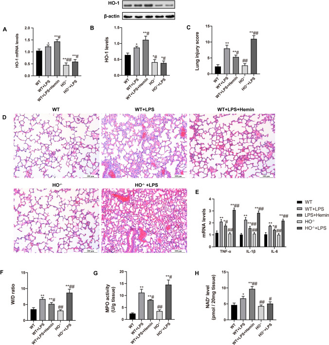
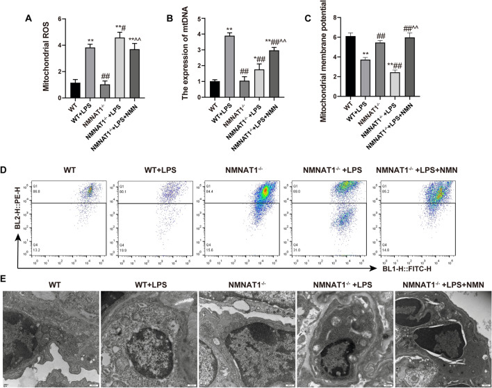
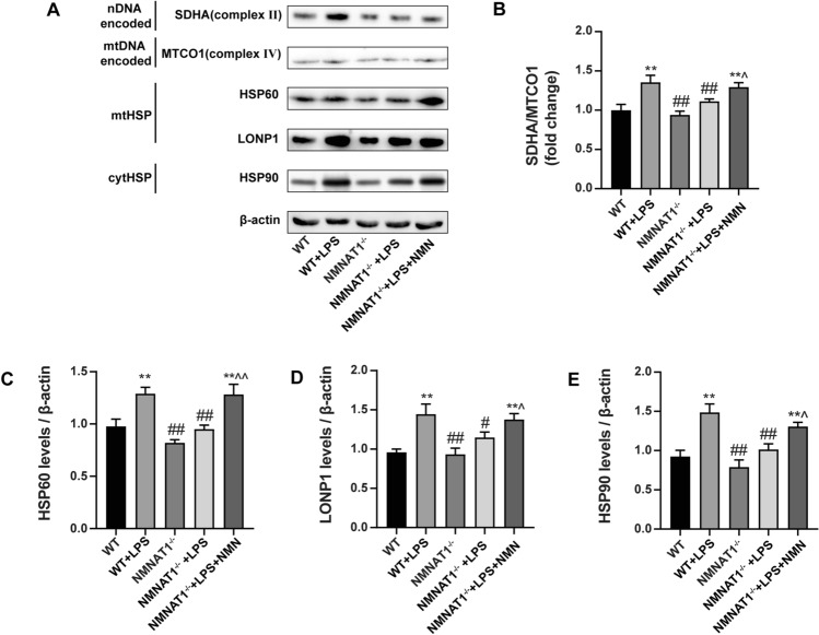
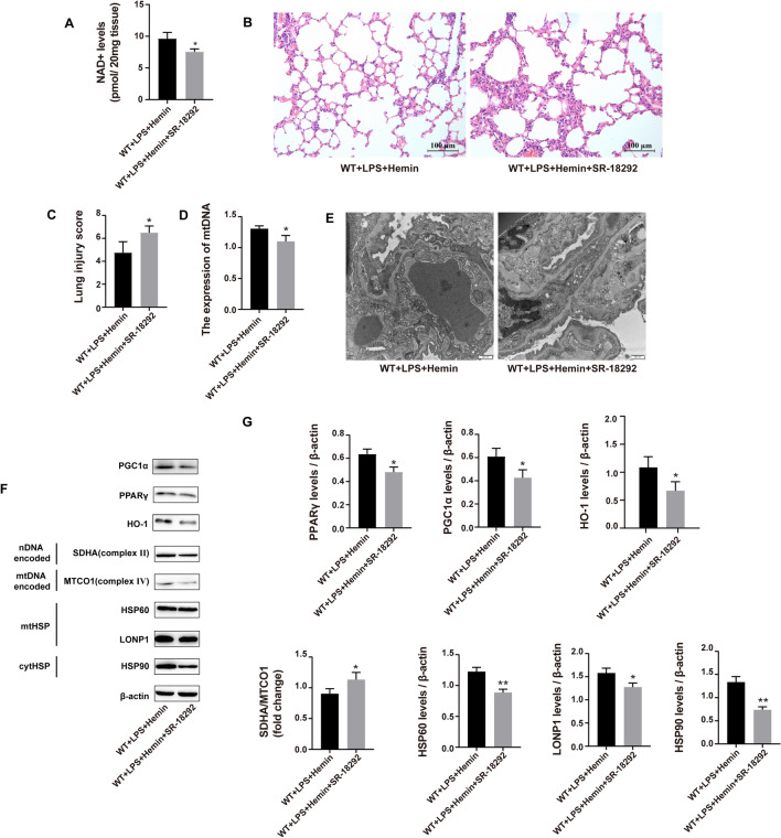
Endotoxin-induced acute lung injury (ALI) is a challenging life-threatening disease for which no specific therapy exists. Mitochondrial dysfunction is corroborated as hallmarks in sepsis which commonly disrupt mitochondria-centered cellular communication networks, especially mitonuclear crosstalk, where the ubiquitous cofactor nicotinamide adenine dinucleotide (NAD+) is essential for mitonuclear communication. Heme oxygenase-1 (HO-1) is critical for maintaining mitochondrial dynamic equilibrium and regulating endoplasmic reticulum (ER) and Golgi stress to alleviating acute lung injury. However, it is unclear whether HO-1 regulates NAD+-mediated mitonuclear communication to exert the endogenous protection during endotoxin-induced ALI. In this study, we observed HO-1 attenuated endotoxin-induced ALI by regulated NAD+ levels and NAD+ affected the mitonuclear communication, including mitonuclear protein imbalance and UPRmt to alleviate lung damage. We also found the protective effect of HO-1 depended on NAD+ and NAD+-mediated mitonuclear communication. Furtherly, the inhibition of the PGC1α/PPARγ signaling exacerbates the septic lung injury by reducing NAD+ levels and repressing the mitonuclear protein imbalance and UPRmt. Altogether, our study certified that HO-1 ameliorated endotoxin-induced acute lung injury by regulating NAD+ and NAD+-mediated mitonuclear communications through PGC1α/PPARγ pathway. The present study provided complementary evidence for the cytoprotective effect of HO-1 as a potential target for preventing and attenuating of endotoxin-induced ALI.
Keywords: Acute lung injury; Heme oxygenase-1; Mitonuclear communication; NAD+; Sepsis.
© 2022. The Author(s), under exclusive licence to Springer Nature Switzerland AG.
Conflict of interest statement
No conflicts of interest are declared by the authors.
Figures


Similar articles
-
Nicotinamide mononucleotide ameliorates acute lung injury by inducing mitonuclear protein imbalance and activating the UPRmt.Exp Biol Med (Maywood). 2022 Jul;247(14):1264-1276. doi: 10.1177/15353702221094235. Epub 2022 May 10. Exp Biol Med (Maywood). 2022. PMID: 35538652 Free PMC article.
-
PI3K/Akt pathway-mediated HO-1 induction regulates mitochondrial quality control and attenuates endotoxin-induced acute lung injury.Lab Invest. 2019 Dec;99(12):1795-1809. doi: 10.1038/s41374-019-0286-x. Epub 2019 Sep 30. Lab Invest. 2019. PMID: 31570770
-
Heme oxygenase-1(HO-1) regulates Golgi stress and attenuates endotoxin-induced acute lung injury through hypoxia inducible factor-1α (HIF-1α)/HO-1 signaling pathway.Free Radic Biol Med. 2021 Mar;165:243-253. doi: 10.1016/j.freeradbiomed.2021.01.028. Epub 2021 Jan 23. Free Radic Biol Med. 2021. PMID: 33493554 Free PMC article.
-
PPARγ-Coactivator-1α, Nicotinamide Adenine Dinucleotide and Renal Stress Resistance.Nephron. 2017;137(4):253-255. doi: 10.1159/000471895. Epub 2017 Jun 8. Nephron. 2017. PMID: 28591759 Free PMC article. Review.
-
Heme oxygenase-1/carbon monoxide: novel therapeutic strategies in critical care medicine.Curr Drug Targets. 2010 Dec;11(12):1485-94. doi: 10.2174/1389450111009011485. Curr Drug Targets. 2010. PMID: 20704552 Review.
Cited by
-
RAGE-TLR4 Crosstalk Is the Key Mechanism by Which High Glucose Enhances the Lipopolysaccharide-Induced Inflammatory Response in Primary Bovine Alveolar Macrophages.Int J Mol Sci. 2023 Apr 10;24(8):7007. doi: 10.3390/ijms24087007. Int J Mol Sci. 2023. PMID: 37108174 Free PMC article.
-
Carbon Dots for the Treatment of Inflammatory Diseases: An Appraisal of In Vitro and In Vivo Studies.Oxid Med Cell Longev. 2023 May 25;2023:3076119. doi: 10.1155/2023/3076119. eCollection 2023. Oxid Med Cell Longev. 2023. PMID: 37273553 Free PMC article. Review.
-
5-Methoxytryptophan ameliorates endotoxin-induced acute lung injury in vivo and in vitro by inhibiting NLRP3 inflammasome-mediated pyroptosis through the Nrf2/HO-1 signaling pathway.Inflamm Res. 2023 Aug;72(8):1633-1647. doi: 10.1007/s00011-023-01769-1. Epub 2023 Jul 17. Inflamm Res. 2023. PMID: 37458783
References
-
- Fowler AA, 3rd, et al. Effect of vitamin C infusion on organ failure and biomarkers of inflammation and vascular injury in patients with sepsis and severe acute respiratory failure: the CITRIS-ALI randomized clinical trial. JAMA. 2019;322:1261–1270. doi: 10.1001/jama.2019.11825. - DOI - PMC - PubMed
MeSH terms
Substances
Grants and funding
LinkOut - more resources
Full Text Sources
