Review
. 2022 Sep 1;323(3):C731-C748.
doi: 10.1152/ajpcell.00210.2022. Epub 2022 Jul 11.

G protein-coupled receptor signaling: transducers and effectors

Affiliations
Review

G protein-coupled receptor signaling: transducers and effectors

Haoran Jiang et al. Am J Physiol Cell Physiol. .

Abstract

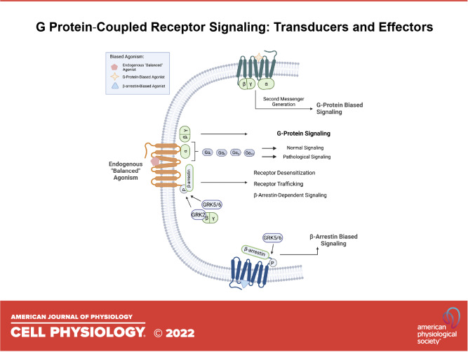
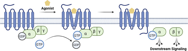
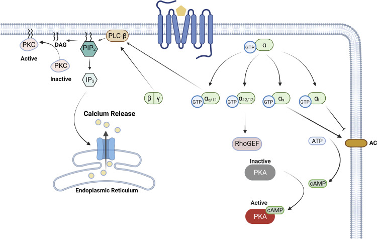
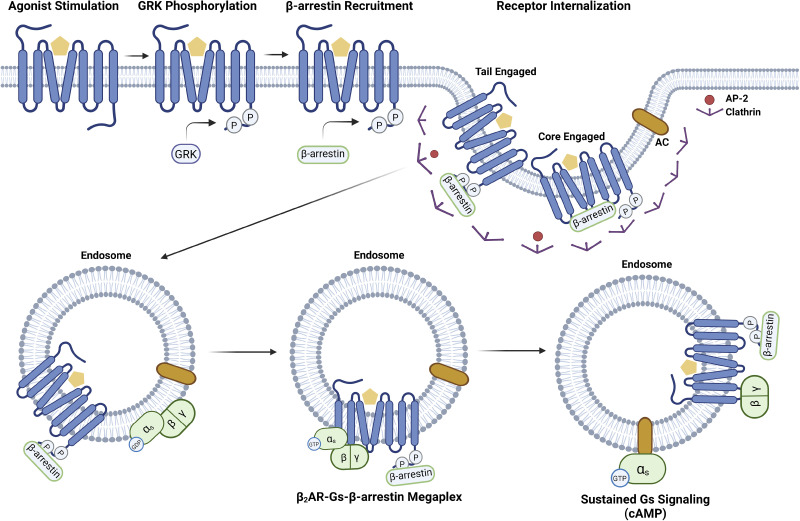
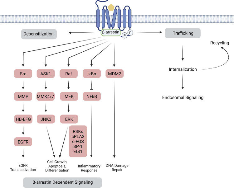
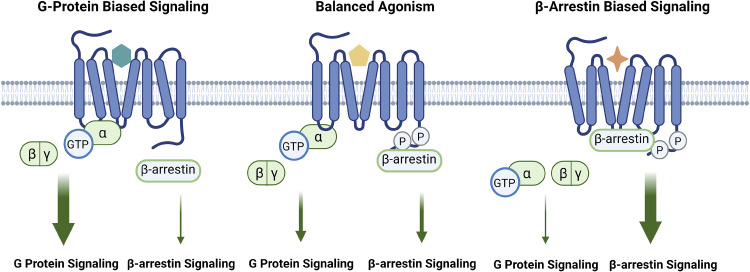
G protein-coupled receptors (GPCRs) are of considerable interest due to their importance in a wide range of physiological functions and in a large number of Food and Drug Administration (FDA)-approved drugs as therapeutic entities. With continued study of their function and mechanism of action, there is a greater understanding of how effector molecules interact with a receptor to initiate downstream effector signaling. This review aims to explore the signaling pathways, dynamic structures, and physiological relevance in the cardiovascular system of the three most important GPCR signaling effectors: heterotrimeric G proteins, GPCR kinases (GRKs), and β-arrestins. We will first summarize their prominent roles in GPCR pharmacology before transitioning into less well-explored areas. As new technologies are developed and applied to studying GPCR structure and their downstream effectors, there is increasing appreciation for the elegance of the regulatory mechanisms that mediate intracellular signaling and function.

Keywords: G protein; G protein-coupled receptor; GPCR kinase; effector; transducer; β-arrestin.


Conflict of interest statement

H. A. Rockman is a scientific cofounder of Trevena Inc., a company that is developing new GPCR ligands. None of the other authors has any conflicts of interest, financial or otherwise, to disclose.

This article is part of the special collection “Advances in GPCRs: Structure, Mechanisms, Disease, and Pharmacology.” Wei Kong, MD, PhD, and Jinpeng Sun, PhD, served as Guest Editors of this collection.

Figures

None
Graphical abstract
Figure 1.
Figure 1.
G protein-coupled receptor activation. Seven-transmembrane GPCRs are a group of diverse receptors in eukaryotes. When an external agonist binds to the GPCR, the receptor will undergo a conformational change, triggering a subsequent interaction with the heterotrimeric G protein complex. The G protein is considered inactive when the α subunit is bound to GDP; however, upon an agonist stimulation, the GDP will physically be displaced by the GTP, rendering the complex active. Both the Gα and the Gβγ subunits will then go on to activate distinct downstream effectors. Figure created with BioRender and published with permission. GPCR, G protein-coupled receptor; GTP, guanosine triphosphate.
Figure 2.
Figure 2.
The heterotrimeric G protein signaling. There are four major families in the α subunit: Gαs, Gαi, Gαq/11, and Gα12/13. Although Gαs activates the AC, Gαi inhibits it, both of which will regulate the intracellular concentration of cAMP. Gαq/11 activates PLC-β, hydrolyzing PIP2 into DAG and IP3. Gα12/13 also has a variety of downstream targets, including RhoGEF. AC, adenyl cyclase; ATP, adenosine triphosphate; cAMP, cyclic adenosine monophosphate; DAG, diacylglycerol; GEF, guanine nucleotide exchange factor; GTP, guanosine triphosphate; IP3, inositol trisphosphate; PIP2, phosphatidylinositol 4,5-bisphosphate; PKA, protein kinase A; PKC, protein kinase C; PLC-β: phospholipase C-β. Figure created with BioRender and published with permission.
Figure 3.
Figure 3.
GPCR desensitization and internalization. Two critical factors are involved in receptor desensitization and internalization after stimulation. The first is GRK phosphorylation of the C-terminal tail of the GPCR. The second is β-arrestin recruitment to the phosphorylated receptor followed by clathrin-dependent internalization. Depending on the GPCR-β-arrestin complex conformation, β-arrestin may either bind to the core (core conformation) or the tail (tail conformation). Because the tail conformation presumably exposes the core for further G protein interactions, it is possible to form a GPCR-G protein-β-arrestin “megaplex” that can exhibit sustained G protein signaling as demonstrated for the β2AR and cytoplasmic cAMP generation (147, 148). Figure created with BioRender and published with permission. β2AR, β2-adrenergic receptor; cAMP, cyclic AMP; GPCR, G protein-coupled receptor; GRK, GPCR kinase.
Figure 4.
Figure 4.
β-Arrestin-mediated signaling. β-arrestins have been shown to regulate a diverse set of signaling pathways involved in a variety of cellular processes. The transducer plays a crucial role in the desensitization, trafficking, internalization, and translocation of its receptor, directing crucial stages of the GPCR life span. β-arrestin has also been shown to activate many signaling cascades, particularly due to its ability to scaffold additional proteins. The wide array of signaling cascades consequently regulate diverse aspects of cellular function, including but not limited to EGFR transactivation, cell growth, apoptosis, cell differentiation, inflammatory responses, and DNA damage repair. ASK1, apoptosis signal-regulating kinase 1; EGFR, epidermal growth factor receptor; ERK, extracellular signal-regulated kinase; GPCR, G protein-coupled receptor; HB-EFG, heparin-binding EGF-like epidermal growth factor; IkBα, NF-κB inhibitor; JNK3, Jun N-terminal kinase 3; MDM2, murine double minute 2 gene (negatively regulates p53); MEK, mitogen-activated protein kinase; MMK4/7, mitogen-activated protein kinase 4/7; MMP, matrix metalloproteinase; NF-κB, nuclear factor-kappa B; RaF, rapidly accelerated fibrosarcoma kinase; Src, protooncogene tyrosine-kinase Src. Figure created with BioRender and published with permission.
Figure 5.
Figure 5.
Biased agonism and signaling selectivity. Biased agonism is a recently emerging concept in which a ligand preferentially activates one signaling pathway over another. In balanced agonism, binding of a ligand potentiates both G protein and β-arrestin signaling. However, when a β-arrestin-biased-ligand binds to a GPCR, conformational changes in the receptor favor β-arrestin-mediated signaling while silencing of G protein-mediated pathways. Similarly, when a G protein-biased ligand binds to the GPCR, conformational changes in the receptor favor G protein-mediated signaling while silencing β-arrestin signaling. Figure created with BioRender.com. GPCR, G protein-coupled receptor; GTP, guanosine triphosphate.

References

    1. Drews J. Paul Ehrlich: magister mundi. Nat Rev Drug Discov 3: 797–801, 2004. doi: 10.1038/nrd1498. - DOI - PubMed
    1. Langley JN. On the reaction of cells and of nerve-endings to certain poisons, chiefly as regards the reaction of striated muscle to nicotine and to curari. J Physiol 33: 374–413, 1905. doi: 10.1113/jphysiol.1905.sp001128. - DOI - PMC - PubMed
    1. Mukherjee C, Caron MG, Coverstone M, Lefkowitz RJ. Identification of adenylate cyclase-coupled β-adrenergic receptors in frog erythrocytes with (minus)-[3-H] alprenolol. J Biol Chem 250: 4869–4876, 1975. doi: 10.1016/s0021-9258(19)41249-0. - DOI - PubMed
    1. Williams LT, Lefkowitz RJ. α-adrenergic receptor identification by (3H)dihydroergocryptine binding. Science 192: 791–793, 1976. doi: 10.1126/science.4894. - DOI - PubMed
    1. Dohlman HG, Caron MG, Lefkowitz RJ. A family of receptors coupled to guanine nucleotide regulatory proteins. Biochemistry 26: 2657–2664, 1987. doi: 10.1021/bi00384a001. - DOI - PubMed

Publication types

MeSH terms