Review
. 2022 Sep:188:114441.
doi: 10.1016/j.addr.2022.114441. Epub 2022 Jul 9.

Protein and polypeptide mediated delivery to the eye

Affiliations
Review

Protein and polypeptide mediated delivery to the eye

Sara Aly Attia et al. Adv Drug Deliv Rev. 2022 Sep.

Abstract

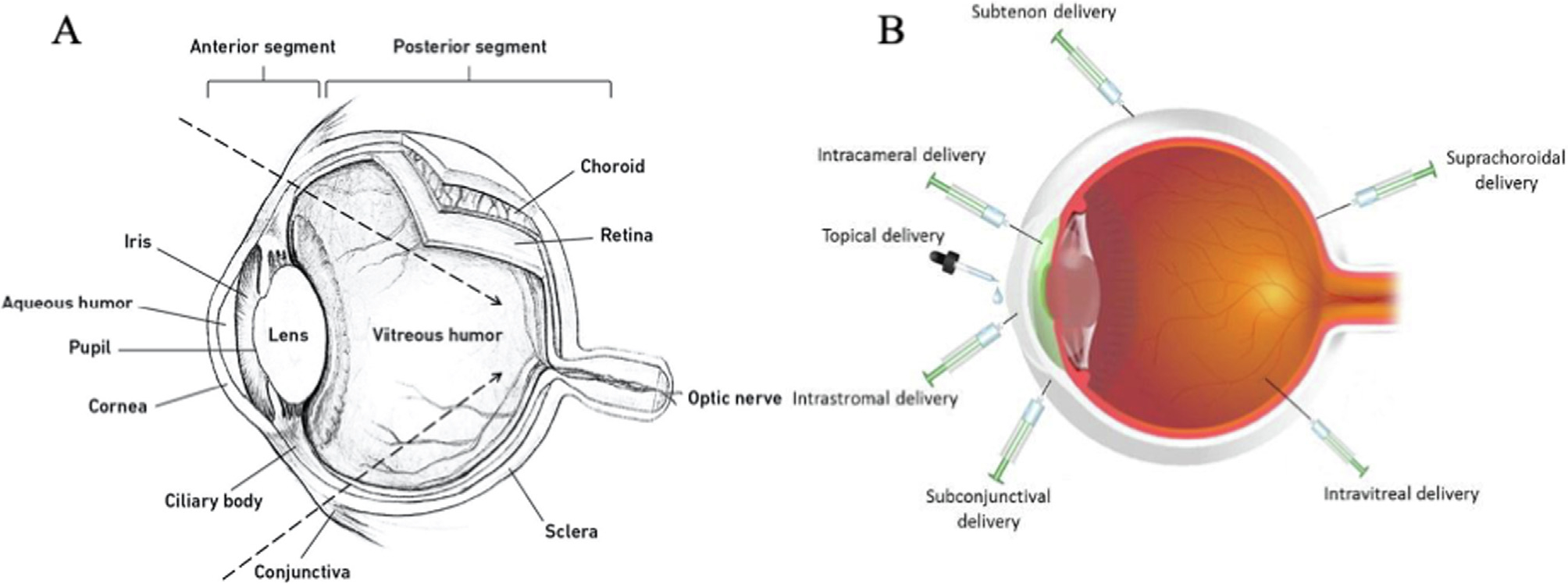
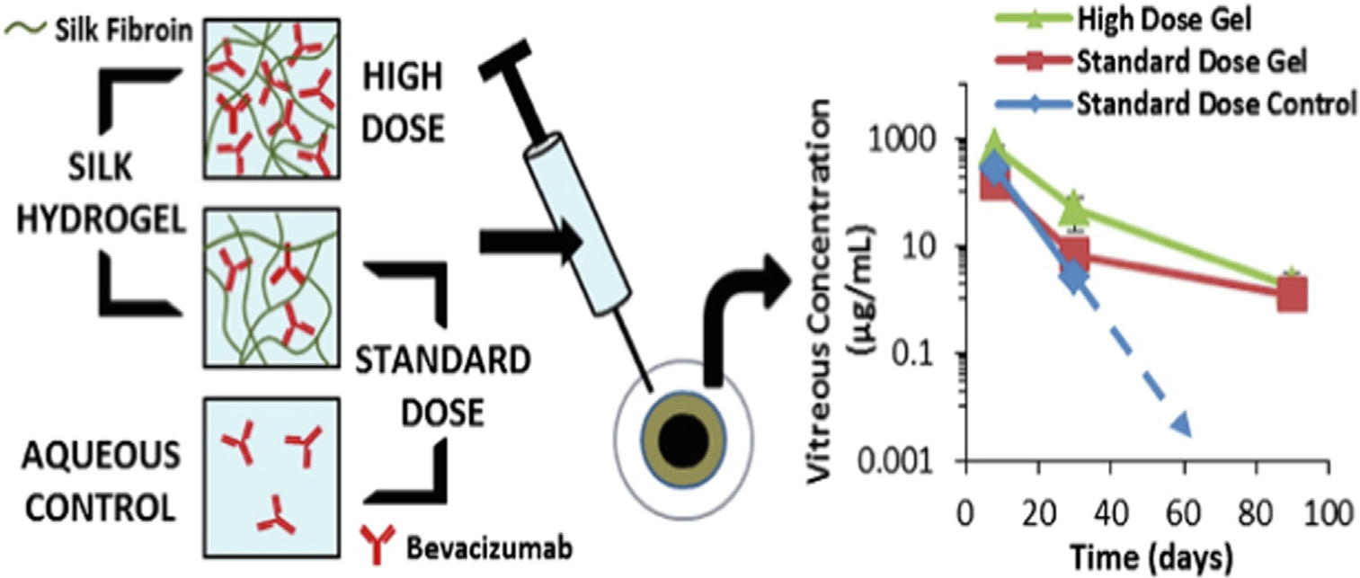
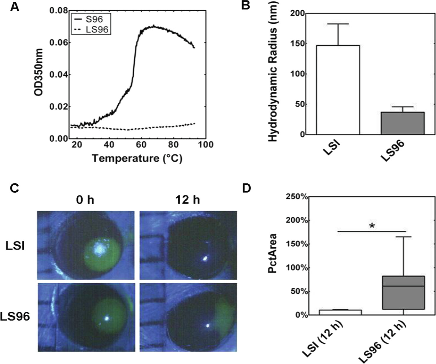
Hybrid or recombinant protein-polymers, peptide-based biomaterials, and antibody-targeted therapeutics are widely explored for various ocular conditions and vision correction. They have been noted for their potential biocompatibility, potency, adaptability, and opportunities for sustained drug delivery. Unique to peptide and protein therapeutics, their production by cellular translation allows their precise modification through genetic engineering. To a greater extent than drug delivery to other systems, delivery to the eye can benefit from the combination of locally-targeted administration and protein-based specificity. Consequently, a range of delivery platforms and administration methods have been exploited to address the ocular delivery of peptide and protein biomaterials. This review discusses a sample of preclinical and clinical opportunities for peptide-based drug delivery to the eye.

Keywords: Antibody; Barriers; Cornea; Elastin-like polypeptides; Lacrimal gland; Ocular; Protein; Subconjunctiva; Vitreous.


Conflict of interest statement

Declaration of Competing Interest The authors declare that they have no known competing financial interests or personal relationships that could have appeared to influence the work reported in this paper.

Figures

Fig. 1.
Fig. 1.
A) Schematic of the ocular anatomy representing the anterior and posterior segments, adapted with permission from [1]. B) Emerging methods for ocular delivery, adapted with permission from [51].
Fig. 2.
Fig. 2.
Functional annotation of the unique proteins identified in the vitreous humor, adapted with permission from the Aretz group [117].
Fig. 3.
Fig. 3.
The reversible phase separation of ELP sustains the retention of rapamycin within the lacrimal gland (LG) of NOD mice. These 12–14 week old male NOD mice have lymphocytic infiltrates in their LGs, dacryoadenitis, and can model dry eye disease characteristic of Sjögren’s Syndrome. Shown here is an example from Ju and colleagues of an ELP fused with FKBP, the cognate binding partner for rapamycin (Rapa). (A) A version called 5FV was developed that forms a depot at body temperature, which is contrasted with a soluble control called 5FA. 5FV contains the more hydrophobic amino acid, valine, in place of alanine residues used in 5FA. Both have similar MW and the capacity to bind 5 Rapa per fusion protein. (B) Following injection into the LG, only 5FV coacervates, which (C) causes long-term drug retention for at least a week after intra-LG injection. (D) As imaged by confocal microscopy of a sectioned LG, the soluble 5FA carrier (red) was cleared from the gland (white) within one day, while (E) the 5FV formulation (red) was retained at high levels. F) A systemic dose of a positive control (FAF-Rapa, Table 1) induced hypercholesteremia in NOD mice, whereas local administration of 5FV-Rapa did not. G) Local 5FV-Rapa increased tear production of the NOD LGs, while systemic FAF-Rapa control could not. The study is adapted with permission from [101]. (For interpretation of the references to color in this figure legend, the reader is referred to the web version of this article.)
Fig. 4.
Fig. 4.
Retina-protective potential of a neuroprotective peptide ‘mini cry’ from the AlphaB crystallin protein fused to an ELP called SI. 129S6/SvEvTac mice were pretreated intravitreally with crySI (Table 1) and the other controls prior to chemical challenge with sodium iodate, NaIO3. (A) Color fundus images of mouse retinas were obtained one week after the challenge. The arrowheads point to the sites of retinal damage, where the scattering of white light enhanced significantly. (B) Image analysis of fundus photography was performed using a white mask, which estimated the percentage of the retinal damage over the entire field (black). (C) Statistical analysis of the retinal degeneration in pretreated mice, by two days, one week, and two weeks, prior to iodate challenge. For all the treatment controls and periods, only crySI showed significant protection of the retina (****p < 0.0001). (D) Histological analysis demonstrated epithelial monolayer disruption in all groups, except in the crySI pretreated groups. Pretreatment with crySI significantly protected the retinal pigment epithelium and neighboring photoreceptors. (E) The pharmacokinetic data showed that the fluorescently labeled crySI and SI had a sustained retention up to 14 days, while mini cry exhibited fast clearance in 3 days. Adapted with permission from [96].
Fig. 5.
Fig. 5.
Schematic representation showing the organization and kinetics profile of drug-loaded silk hydrogels at a standard and high drug dose. As shown, both standard and high dose of bevacizumab achieved equivalent or greater concentrations within the vitreous humor up to three months, compared to the positive control that fell below the quantification after 1 month, adapted with permission from [149].
Fig. 6.
Fig. 6.
ELP-mediated assembly enhances lacritin-mediated closure of a defect in the corneal endothelium. (A) Optical density (OD) demonstrates that LS96 lacks a detectable phase separation across a wide range of temperatures, in contrast to S96 ELP alone (shown) and LSI, which assembles larger nanostructures above ~ 19 °C (data not shown). (B) Dynamic light scattering (DLS) confirms that LSI forms nanoparticles with a larger hydrodynamic radius, Rh, compared to LS96 at 37 °C. (C) A 2 mm corneal abrasion was created on the NOD mouse eye, treated with either LSI or LS96, imaged by fluorescein staining, and quantified by a blinded reviewer. LSI treated corneas had almost undetectable defects by 12 h. (D) While both LSI and LS96 contained the lacritin protein fused to a similar MW ELP, the ELP-mediated assembly of LSI nanoparticles significantly decreased the area of the defect at 12 hrs (PcTArea) (p < 0.05, n = 8). This data is adapted by permission from [104].

Similar articles

Cited by

References

    1. Johannsdottir S, Jansook P, Stefansson E, Kristinsdottir IM, Fulop Z, Asgrimsdottir GM, et al., Topical drug delivery to the posterior segment of the eye: Dexamethasone concentrations in various eye tissues after topical administration for up to 15 days to rabbits, J. Drug Delivery Sci. Technol. 45 (2018) 449–454.
    1. Gorantla S, Rapalli VK, Waghule T, Singh PP, Dubey SK, Saha RN, et al., Nanocarriers for ocular drug delivery: current status and translational opportunity, RSC Adv. 10 (46) (2020) 27835–27855. - PMC - PubMed
    1. Rodrigues GA, Lutz D, Shen J, Yuan X, Shen H, Cunningham J, et al., Topical drug delivery to the posterior segment of the eye: addressing the challenge of preclinical to clinical translation, Pharm. Res. 35 (12) (2018) 245. - PMC - PubMed
    1. Cholkar K, Dasari SR, Pal D, Mitra AK, 1 - Eye: anatomy, physiology and barriers to drug delivery, in: Mitra AK (Ed.), Ocular Transporters and Receptors, Woodhead Publishing, 2013, pp. 1–36.
    1. Joseph RR, Venkatraman SS, Drug delivery to the eye: what benefits do nanocarriers offer?, Nanomedicine. 12 (6) (2017) 683–702. - PubMed

Publication types