. 2022 Sep:171:105814.
doi: 10.1016/j.nbd.2022.105814. Epub 2022 Jul 8.

Mild hypothermia fails to protect infant macaques from brain injury caused by prolonged exposure to Antiseizure drugs

Affiliations

Mild hypothermia fails to protect infant macaques from brain injury caused by prolonged exposure to Antiseizure drugs

Chrysanthy Ikonomidou et al. Neurobiol Dis. 2022 Sep.

Abstract

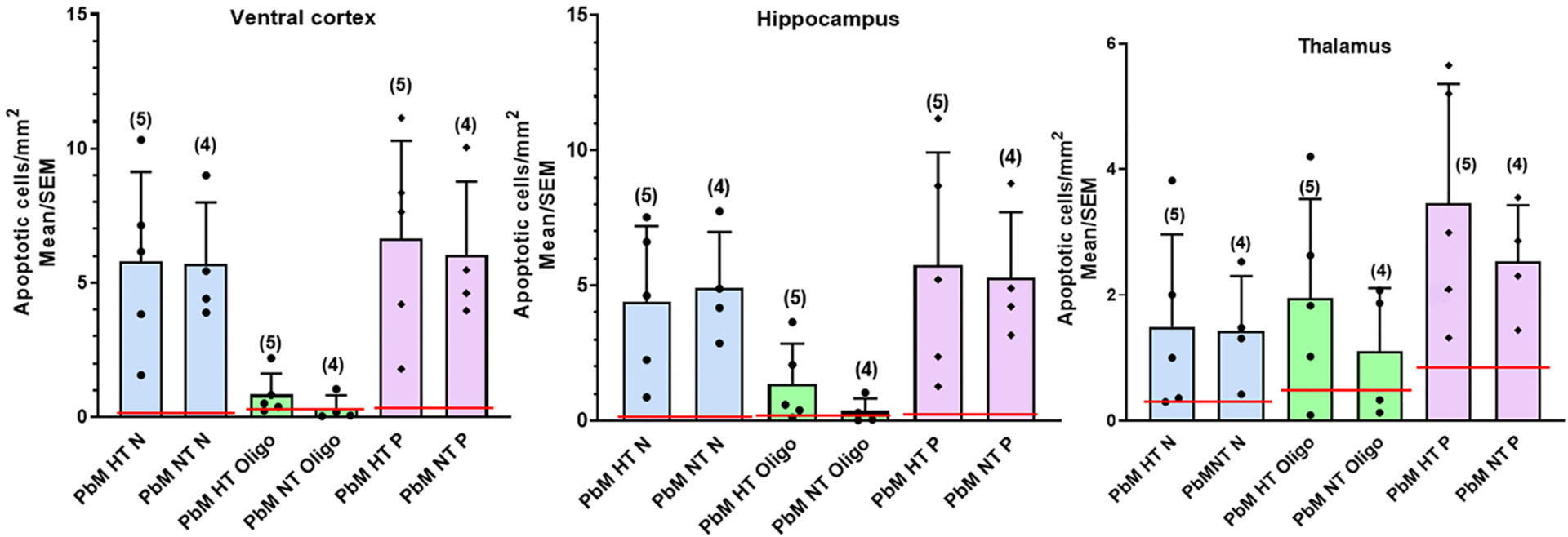
Barbiturates and benzodiazepines are GABAA-receptor agonists and potent antiseizure medications. We reported that exposure of neonatal macaques to combination of phenobarbital and midazolam (Pb/M) for 24 h, at clinically relevant doses and plasma levels, causes widespread apoptosis affecting neurons and oligodendrocytes. Notably, the extent of injury was markedly more severe compared to shorter (8 h) exposure to these drugs. We also reported that, in the infant macaque, mild hypothermia ameliorates the apoptosis response to the anesthetic sevoflurane. These findings prompted us explore whether mild hypothermia might protect infant nonhuman primates from neuro- and gliotoxicity of Pb/M. Since human infants with seizures may receive combinations of benzodiazepines and barbiturates for days, we opted for 24 h treatment with Pb/M. Neonatal rhesus monkeys received phenobarbital intravenously, followed by midazolam infusion over 24 h under normothermia (T > 36.5 °C-37.5 °C; n = 4) or mild hypothermia (T = 35 °C-36.5 °C; n = 5). Medication doses and blood levels measured were comparable to those in human infants. Animals were euthanized at 36 h and brains examined immunohistochemically and stereologically. Treatment was well tolerated. Extensive degeneration of neurons and oligodendrocytes was seen at 36 h in both groups within neocortex, basal ganglia, hippocampus and brainstem. Mild hypothermia over 36 h (maintained until terminal perfusion) conferred no protection against the neurotoxic and gliotoxic effects of Pb/M. This is in marked contrast to our previous findings that mild hypothermia is protective in the context of a 5 h-long exposure to sevoflurane in infant macaques. These findings demonstrate that brain injury caused by prolonged exposure to Pb/M in the neonatal primate cannot be ameliorated by mild hypothermia.

Keywords: Antiseizure; Apoptosis; Barbiturate; Benzodiazepine; Brain injury; Development; Sedative.


Figures

Fig. 1.
Fig. 1.
Apoptosis in the neonatal rhesus macaque brain following Pb/M exposure under normothermic and hypothermic conditions. A - C show computer generated plots of neuroapoptosis (red dots) and oligoapoptosis (green dots) in the brains of a rhesus control infant (left), a normothermic infant exposed to Pb/M for 24 h and euthanized at 36 h (middle) and a hypothermic infant exposed to Pb/M for 36 h and euthanized at 36 h (right). There is substantial amount of neuro- and oligoapoptosis in the brains of both treated infants. At this level and at both time points there is homogeneous pattern of neuro and oligoapoptosis in the caudate, extending into the thalamus, hypothalamus, corpus callosum and subcortical white matter. A laminar pattern of neuronal apoptosis appears within the cingulate, frontal somatosensory, insular, temporal, entorhinal cortices and subiculum. There is no apparent difference in the amount of injury when comparing the normothermic and hypothermic infants. Scale bar = 5 mm.
Fig. 2.
Fig. 2.
Light micrographs depicting AC3 positive profiles (red arrows) within the insular cortex in a normothermic rhesus infant exposed to Pb/M (A) and a hypothermic infant exposed to the drug combination +36 h of hypothermia (D). Perfusion fixation occurred for both animals at 36 h. Note that AC3 immuno-positive neurons are present at high density in layer II of the cortex and that neuronal processes are prominently stained. B, C, E and F show magnified views of the black rectangles in A and D.
Fig. 3.
Fig. 3.
Apoptosis induced by Pb/M affects neurons and oligodendrocytes. Composites A-C show confocal images of NeuN, MBP, GFAP, AC3 and DAPI stained sections. There is colocalization of NeuN with AC3 and MBP with AC3 indicating neuronal and oligodendroglia apoptosis respectively. In C, GFAP staining (green) does not colocalize with AC3 (red) indicating that astroglia are not affected by the proapoptotic effect of Pb/M.
Fig. 4.
Fig. 4.
Mild hypothermia does not protect from apoptosis induced by Pb/M in the neonatal macaque brain. Pb/M induces widespread apoptosis in the infant macaque brain affecting primarily neurons at 36 h. The graphs illustrate numbers of apoptotic neurons (blue), apoptotic oligodendrocytes (green) and apoptotic profiles (purple) counted in the brains of infants treated with Pb/M under normothermic (NT: n = 4) or hypothermic (HT: n = 5) conditions and euthanized at 36 h. The red lines demonstrate mean numbers of apoptosis reported previously in control animals for each cell type (Noguchi et al., 2021; N: 95,476 ± 21,807; Oligo: 247,372 ± 81,510; P: 342,849 ± 96,592; n = 5). Columns represent means ± SEM of total numbers of neurons (N; blue), oligodendrocytes (Oligo; green) and total apoptotic profiles (P; purple), counted in the whole brain. Statistical comparisons between NT and HT groups were performed by means of Student’s test-test. There were no statistically significant differences between the two groups. Abbreviations: Pb/M: phenobarbital/midazolam; HT: hypothermia; NT: normothermia; N: neurons; Oligo: oligodendrocytes; P: profiles.
Fig. 5.
Fig. 5.
Effects of Pb/M at 36 h on apoptosis of neurons (N), oligodendrocytes (Oligo) and total profiles (P: N + Oligo) in the ventral cortex, hippocampus and thalamus under normothermic (NT) and hypothermic (HT) conditions. Within the ventral cortex and the hippocampus, Pb/M exposure had a significant effect on neuronal apoptosis (blue columns) and total profiles (purple) but not on oligoapoptosis (green columns). Hypothermia did not protect from neuro- or oligoapoptosis. Columns represent means ± SEM of densities (cells per square millimeter) of neurons (blue), oligodendrocytes (Oligos; green) and total apoptotic profiles (purple). Numbers in parentheses represent numbers of animals in each group. The red lines depict mean densities previously reported in control animals (Noguchi et al., 2021: Ventral Cortex: N: 0.104 ± 0.019; Oligo: 0.140 ± 0.040; P: 0.246 ± 0.043; Hippocampus: N: 0.156 ± 0.042; Oligo 0.138 ± 0.042; P: 0.292 ± 0.049; Thalamus: N: 0.400 ± 0.144; Oligo: 0.484 ± 0.234; P: 0.886 ± 0.253; n = 5). No significant differences were detected for any cell type between the normothermia and hypothermia groups (Student’s t-test). Abbreviations: Pb/M: phenobarbital/midazolam; HT: hypothermia; NT: normothermia; N: neurons; Oligo: oligodendrocytes; P: profiles.

Similar articles

Cited by

References

    1. Aihara H, Okada Y, Tamaki N, 2001. The effects of cooling and rewarming on the neuronal activity of pyramidal neurons in guinea pig hippocampal slices. Brain Res. 893, 36–45. - PubMed
    1. Bittigau P, Sifringer M, Genz K, et al., 2002. Antiepileptic drugs and apoptotic neurodegeneration in the developing brain. Proc. Natl. Acad. Sci 99, 15089–15094. - PMC - PubMed
    1. Brambrink AM, Evers AS, Avidan MS, et al., 2010. Isoflurane-induced neuroapoptosis in the neonatal rhesus macaque brain. Anesthesiology 112, 834–841. - PMC - PubMed
    1. Brambrink AM, Back SA, Riddle A, et al., 2012. Isoflurane-induced apoptosis of oligodendrocytes in the neonatal primate brain. Ann. Neurol 2, 525–535. - PMC - PubMed
    1. Creeley CE, Olney JW, 2010. The young: neuroapoptosis induced by anesthetics and what to do about it. Anesth. Analg 110, 442–448. - PubMed

Publication types