. 2022 Nov:109:109102.
doi: 10.1016/j.jnutbio.2022.109102. Epub 2022 Jul 8.

N-3 polyunsaturated fatty acids block the trimethylamine-N-oxide- ACE2- TMPRSS2 cascade to inhibit the infection of human endothelial progenitor cells by SARS-CoV-2

Affiliations

N-3 polyunsaturated fatty acids block the trimethylamine-N-oxide- ACE2- TMPRSS2 cascade to inhibit the infection of human endothelial progenitor cells by SARS-CoV-2

En-Pei Isabel Chiang et al. J Nutr Biochem. 2022 Nov.

Abstract

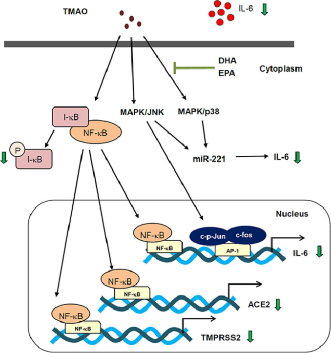
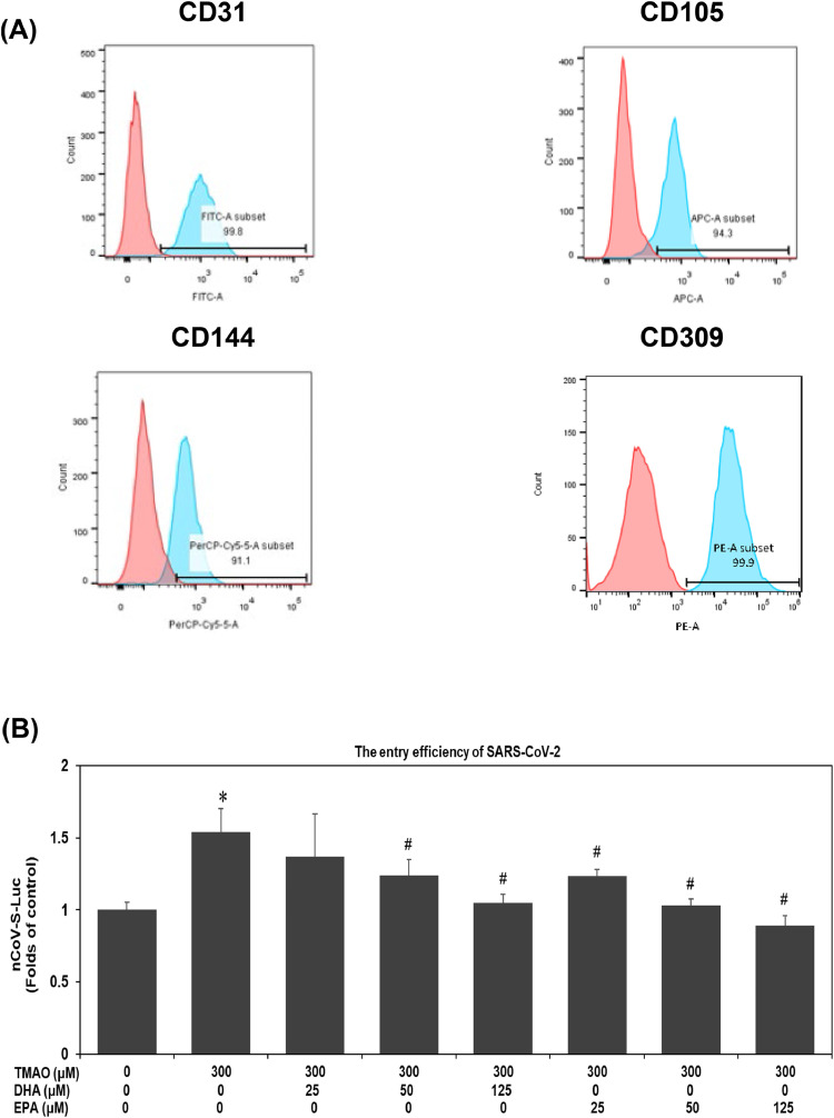
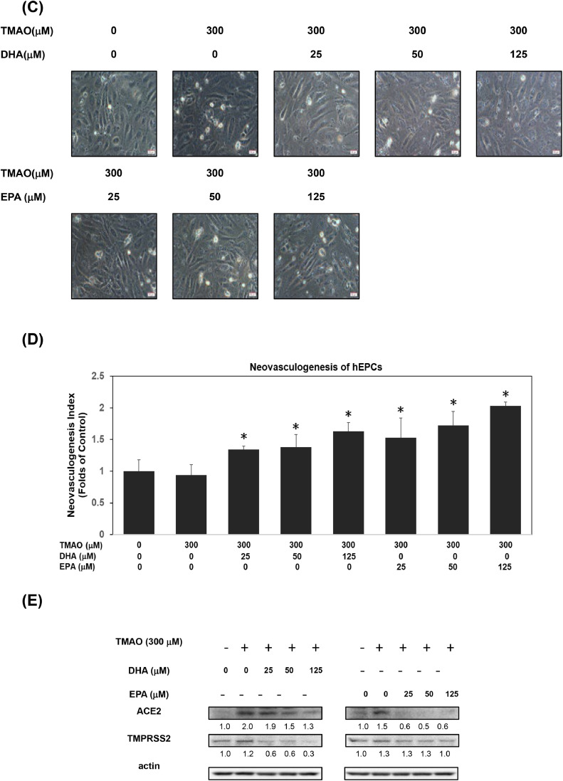
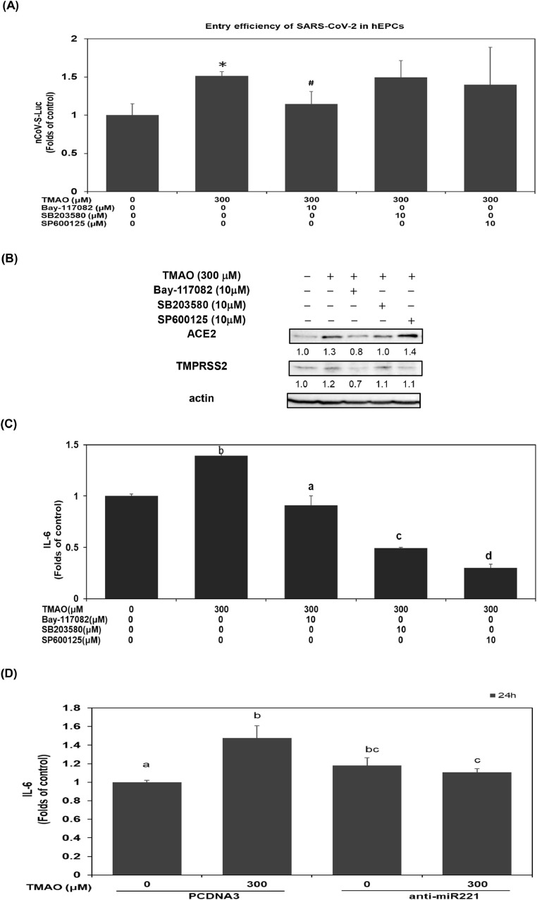
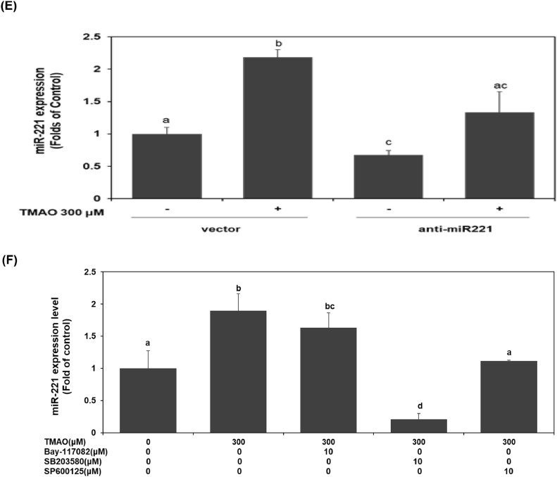
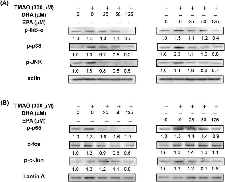
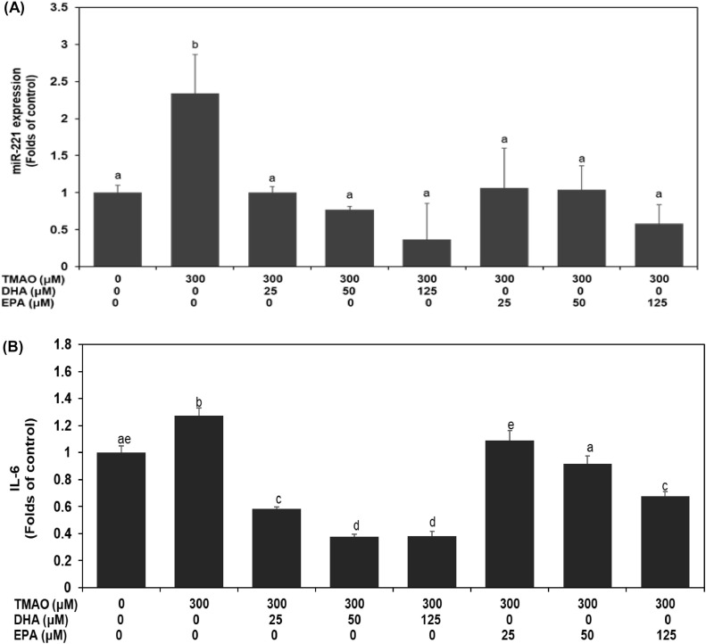
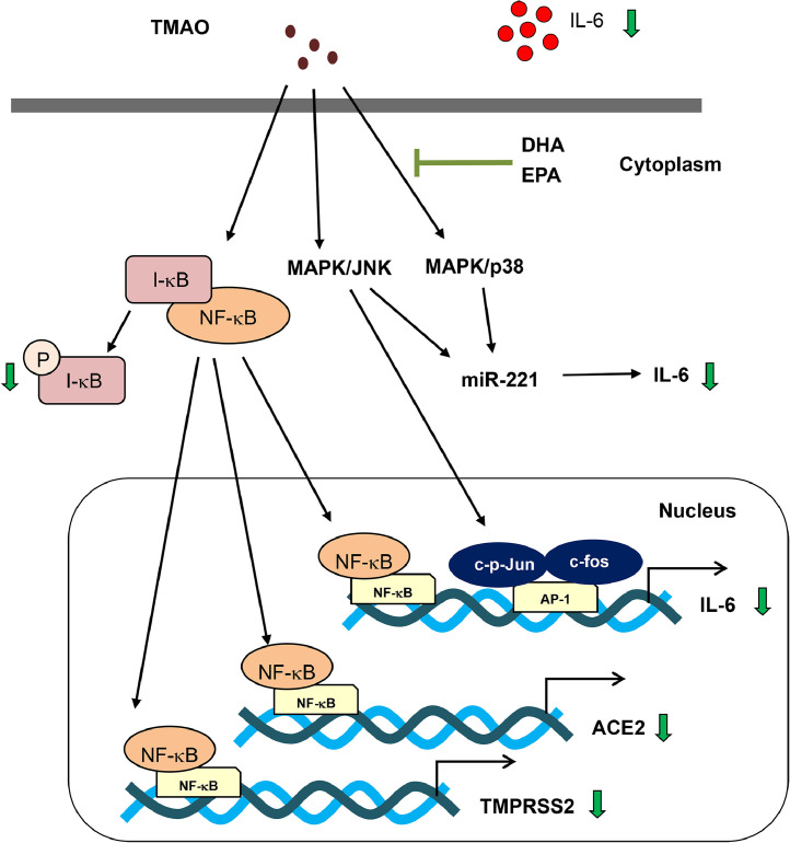
Severe acute respiratory syndrome coronavirus 2(SARS-CoV-2) is a novel coronavirus that infects many types of cells and causes cytokine storms, excessive inflammation, acute respiratory distress to induce failure of respiratory system and other critical organs. In this study, our results showed that trimethylamine-N-oxide (TMAO), a metabolite generated by gut microbiota, acts as a regulatory mediator to enhance the inerleukin-6 (IL-6) cytokine production and the infection of human endothelial progenitor cells (hEPCs) by SARS-CoV-2. Treatment of N-3 polyunsaturated fatty acids (PUFAs) such as docosahexaenoic acid (DHA) and eicosapentaenoic acid (EPA) could effectively block the entry of SARS-CoV-2 in hEPCs. The anti-infection effects of N-3 PUFAs were associated with the inactivation of NF-κB signaling pathway, a decreased expression of the entry receptor angiotensin-converting enzyme 2 (ACE2) and downstream transmembrane serine protease 2 in hEPCs upon the stimulation of TMAO. Treatment of DHA and EPA further effectively inhibited TMAO-mediated expression of IL-6 protein, probably through an inactivation of MAPK/p38/JNK signaling cascades and a downregulation of microRNA (miR)-221 in hEPCs. In conclusion, N-3 PUFAs such as DHA and EPA could effectively act as preventive agents to block the infection of SARS-CoV-2 and IL-6 cytokine production in hEPCs upon the stimulation of TMAO.

Keywords: SARS-CoV-2; Trimethylamine-N-oxide; human endothelial progenitor cells; interleukin-6; microRNA-221.


Figures

Image, graphical abstract
Graphical abstract
Fig 1
Fig. 1
N-3 PUFAs effectively block the entry of SARS-CoV-2 and the expression of ACE2 and TMPRSS2 proteins in hEPCs upon TMAO stimulation (A)Characterization of specific cell surface markers in hEPCs by flow cytometry analysis. hEPCs were cultured with TMAO (0 and 300 μM) in the presence of DHA and EPA (at concentrations of 0, 25, 50, and 125 μM, respectively) for 24 h. (B) Measurement of entry efficacy was performed by using SARS-CoV-2 plasmid (nCoV-S-Luc) infection as described in Materials and Methods. Experiments were carried out in triplicate. The values are presented as mean ± standard deviation (SD) in each subgroup. An asterisk represented a statistical difference in comparison with untreated control subgroup (P<.05). A pound sign represented a statistical difference in comparison with TMAO (300 μM) -treated subgroup (P<.05). (C) At the end of SARS-CoV-2 plasmid transfection, photomicrographs of cell morphology were also documented by using inverted phase-contrast microscope (300X). In the lower right corner of each images, a scale bar represented 30 μM (D) Neovasculogenesis levels of SARS-CoV-2 plasmid infected hEPCs were measured at 6 h time point. A detailed neovasculogenesis procedure was described in Materials and Methods Section. An asterisk represented a statistical difference in comparison with TMAO-treated alone subgroup (P<.05). (E) Measurement of cytoplasmic proteins including ACE2, TMPRSS2 and actin was performed by using Western Blotting analysis as described in Materials and Methods. The integrated densities of each protein (ACE2 and TMPRSS2) were adjusted with the corresponding internal control protein (actin) and shown in the bottom row.
Fig 2
Fig. 2
TMAO mediated the infection of hEPCs by SARS-CoV-2 and the expression of IL-6 protein through modulation of cellular signaling pathways and microRNA-221 level. hEPCs were cultured with TMAO (0 and 300 μM) in the presence or absence of Bay-117082, SB203580, or SP600125 (at a concentration of 10 μM) for 24 h. (A) Measurement of entry efficacy was performed by using SARS-CoV-2 plasmid (nCoV-S-Luc) infection as described in Materials and Methods. Experiments were carried out in triplicate. The values are presented as mean ± SD in each subgroup. An asterisk represented a statistical difference in comparison with untreated control subgroup (P<.05). A pound sign represented a statistical difference in comparison with TMAO (300 μM)-treated subgroup (P<.05). (B) Measurement of cytoplasmic proteins including ACE2, TMPRSS2, and actin was performed by using Western Blotting analysis as described in Materials and Methods. The integrated densities of each protein (ACE2 and TMPRSS2) were adjusted with the corresponding internal control proteins (actin) and shown in the bottom row. Measurement of IL-6 protein (C) and miR-221 (F) and was performed by using ELISA assay and qPCR analysis, respectively, as described in Materials and Methods. (D, E) hEPCs were cultured with TMAO (0 and 300 μM) in the presence or absence of anti-sense plasmid against miR-221 (anti-miR-221) for 24 h. Measurement of IL-6 protein (D) and miR-221 (E) was performed as described in Materials and Methods. Different letters represent statistical differences among different subgroups (P<.05).
Fig 3
Fig. 3
N-3 PUFAs effectively blocked TMAO-mediated activation of signaling pathways in hEPCs hEPCs were cultured with TMAO (0 and 300 μM) in the presence or absence of DHA and EPA (at concentrations of 0, 25, 50, and 125 μM) for 24 h. Measurement of (A) cytoplasmic proteins (p-IκB-α, p-p38, p-JNK, and actin) and (B) nuclear proteins (p-p65, c-fos, p-c-Jun, and lamin A) was performed by using Western Blotting analysis as described in Materials and Methods. The integrated densities of each cytoplasmic proteins (p-IκB-α, p-p38, p-JNK) and nuclear proteins (p-p65, c-fos, p-c-Jun) were adjusted with the corresponding control proteins actin and lamin A, respectively. The quantification values were shown in the bottom row.
Fig 4
Fig. 4
N-3 PUFAs significantly inhibit TMAO-mediated expression of IL-6 protein through downregulation of miR-221 in hEPCs hEPCs were cultured with TMAO (0 and 300 μM) in the presence or absence of DHA and EPA (at concentrations of 0, 25, 50, and 125 μM) for 24 h. Measurement of miR-221 (A) and IL-6 protein (B) was performed by using qPCR analysis and ELISA assay, respectively, as described in Materials and Methods. Different letters represent statistical differences among different subgroups (P<.05).
Fig 5
Fig. 5
Proposed mechanisms of signaling pathways regulated by N-3 PUFAs in hEPCs upon TMAO stimulation. Green arrows indicate decreases in expression level.

Similar articles

Cited by

References

    1. Harcourt J, Tamin A, Lu X, Kamili S, Sakthivel SK, Murray J, et al. Severe Acute Respiratory Syndrome Coronavirus 2 from patient with coronavirus disease, United States. Emerg Infect Dis. 2020;26:1266–1273. - PMC - PubMed
    1. Furukawa NW, Brooks JT, Sobel J. Evidence supporting transmission of severe acute respiratory syndrome Coronavirus 2 while presymptomatic or asymptomatic. Emerg infect dis. 2020;26:e1–e6. - PMC - PubMed
    1. Shahid Z, Kalayanamitra R, McClafferty B, Kepko D, Ramgobin D, Patel R, et al. COVID-19 and older adults: what we know. J Am Geriatrics Soc. 2020;68:926–929. - PMC - PubMed
    1. Zabetakis I, Lordan R, Norton C, Tsoupras A. COVID-19: the inflammation link and the role of nutrition in potential mitigation. Nutrients. 2020;12(5):1466. - PMC - PubMed
    1. Butler MJ, Barrientos RM. The impact of nutrition on COVID-19 susceptibility and long-term consequences. Brain, behav, and immun. 2020;87:53–54. - PMC - PubMed

Publication types