. 2022:150:25-89.
doi: 10.1016/bs.ctdb.2022.03.003. Epub 2022 Jun 11.

Receptor control by membrane-tethered ubiquitin ligases in development and tissue homeostasis

Affiliations

Receptor control by membrane-tethered ubiquitin ligases in development and tissue homeostasis

Andres M Lebensohn et al. Curr Top Dev Biol. 2022.

Abstract

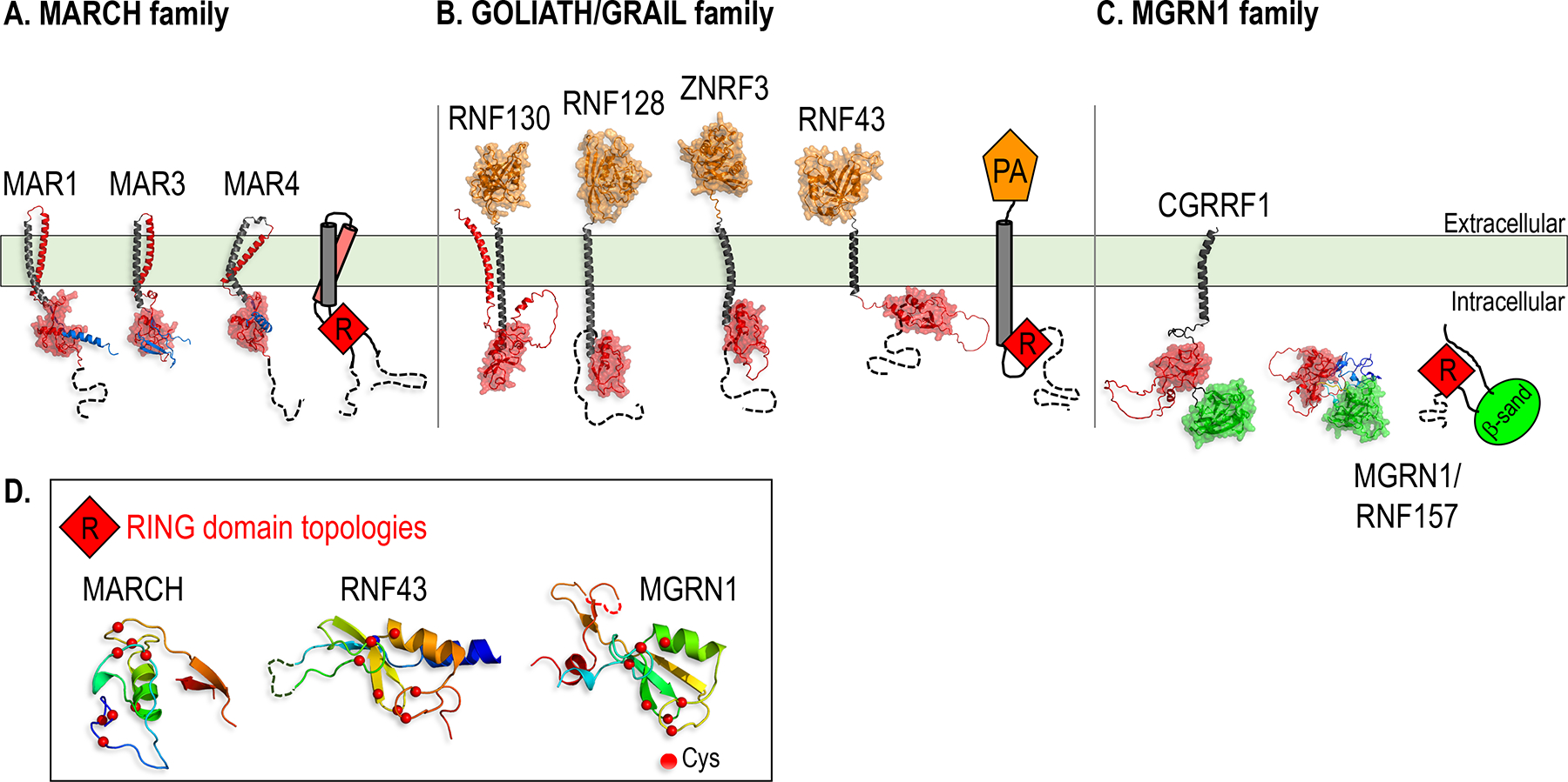
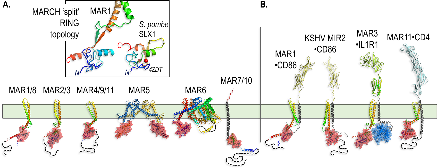
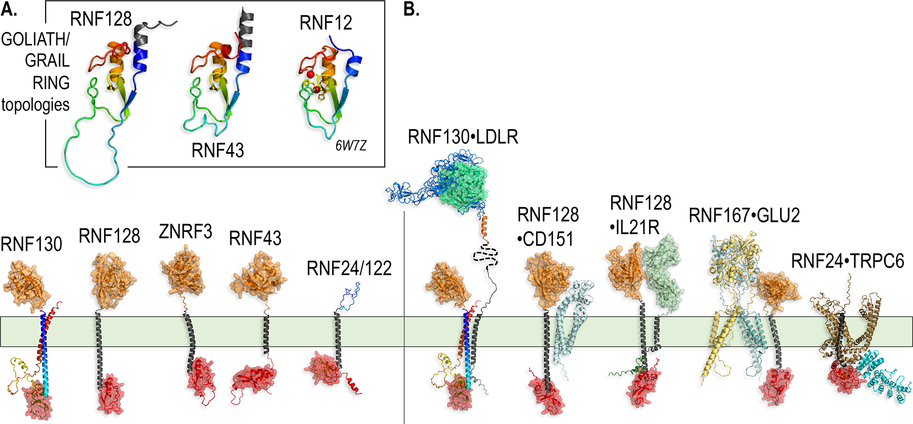
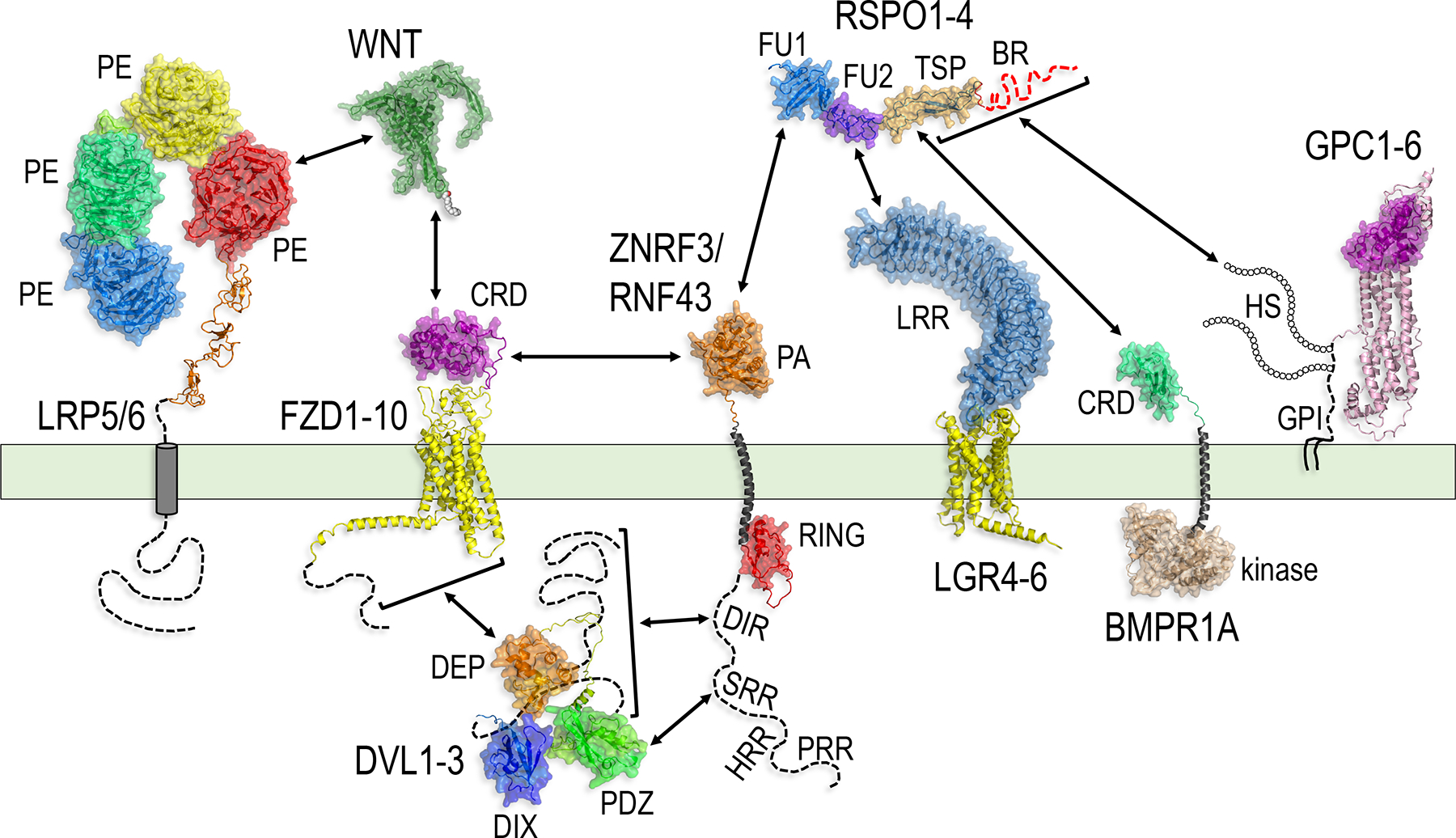
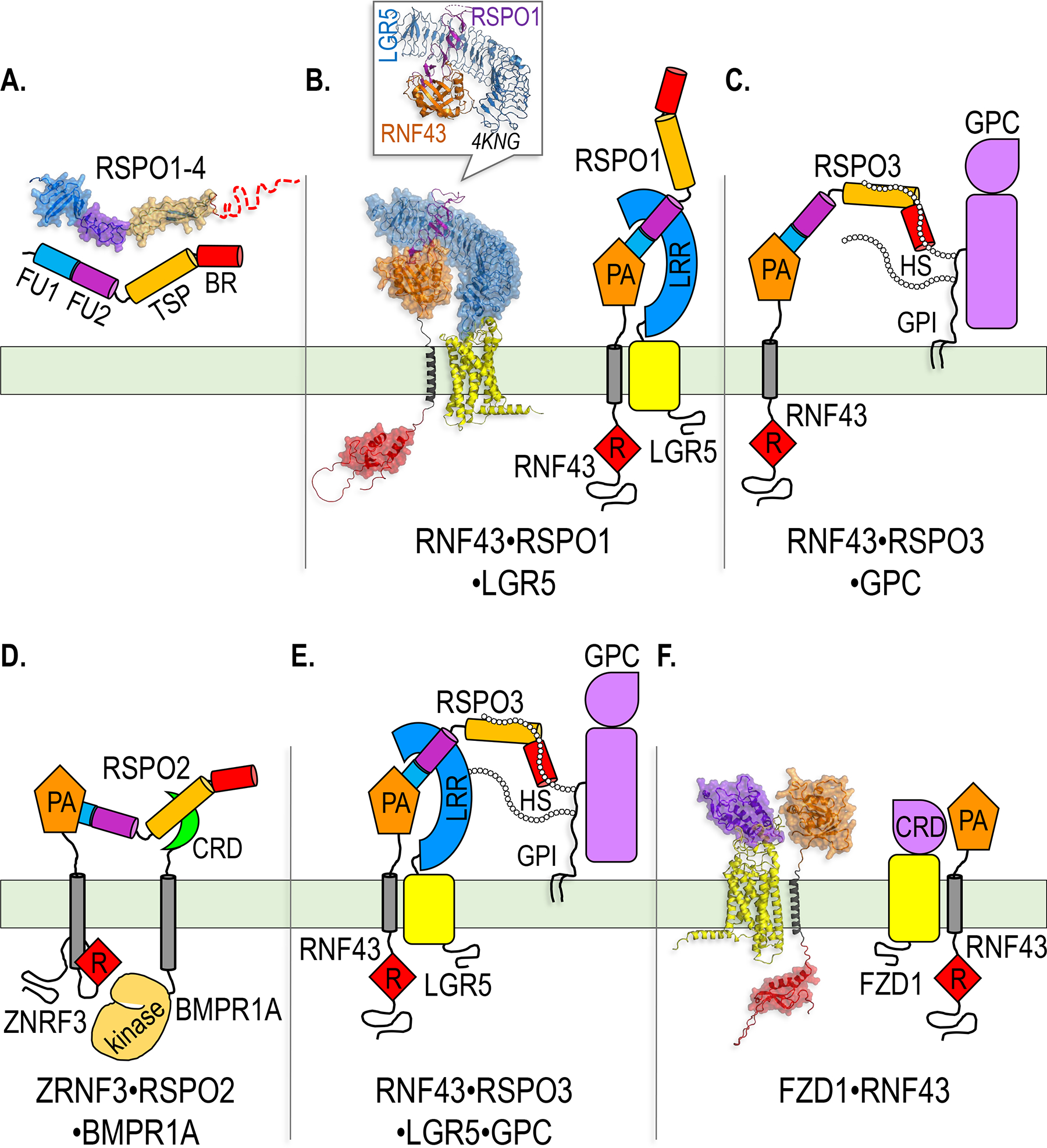
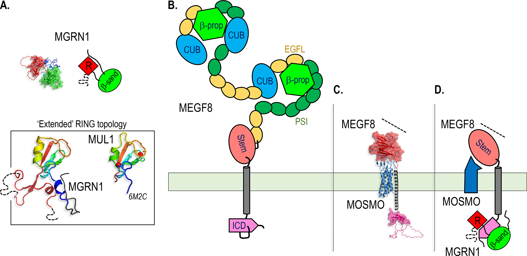
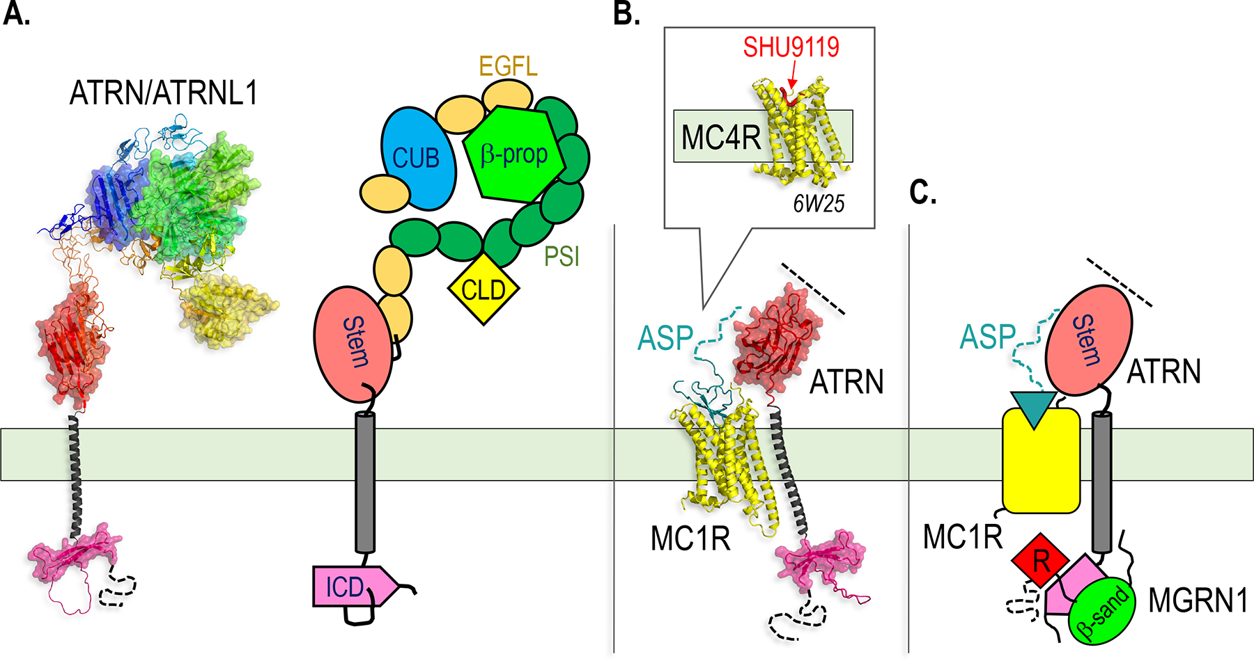
Paracrine cell-cell communication is central to all developmental processes, ranging from cell diversification to patterning and morphogenesis. Precise calibration of signaling strength is essential for the fidelity of tissue formation during embryogenesis and tissue maintenance in adults. Membrane-tethered ubiquitin ligases can control the sensitivity of target cells to secreted ligands by regulating the abundance of signaling receptors at the cell surface. We discuss two examples of this emerging concept in signaling: (1) the transmembrane ubiquitin ligases ZNRF3 and RNF43 that regulate WNT and bone morphogenetic protein receptor abundance in response to R-spondin ligands and (2) the membrane-recruited ubiquitin ligase MGRN1 that controls Hedgehog and melanocortin receptor abundance. We focus on the mechanistic logic of these systems, illustrated by structural and protein interaction models enabled by AlphaFold. We suggest that membrane-tethered ubiquitin ligases play a widespread role in remodeling the cell surface proteome to control responses to extracellular ligands in diverse biological processes.

Keywords: ATRN; BMP; BMPR1A; Cancer; Cell surface receptor; DVL; Development; E3; Endocytosis; FZD; Frizzled; HSPG; Hedgehog; Heparan sulfate proteoglycan; LGR; LRP6; MARCH; MEGF8; MGRN1; MMM; MOSMO; Melanocortin; Membrane-tethered; Morphogen; PROTAC; Patterning; Primary cilium; Protein degradation; R-spondin; RING; RNF43; RSPO; Regeneration; Signaling; Smoothened; Stem cells; Tissue homeostasis; Ubiquitin ligase; Ubiquitylation; WNT; ZNRF3.


Figures

Figure 1.
Figure 1.
Structural models of the three classes of TM and membrane-associated E3s discussed in this chapter. A-C. AlphaFold models of representative members of the MARCH (A), GOLIATH/GRAIL (B) and MGRN1 (C) E3 families, with cartoons used throughout the figures to represent each family. The RING domain, which recruits a Ub-charged E2 for Ub transfer to the substrate, is colored red and shown with a space filling surface in the structural models, and is shown as a red diamond labeled “R” in the cartoons in this and all subsequent figures. All the structures shown in the figures represent AlphaFold models unless indicated otherwise with a Protein Data Bank (PBD) ID shown in italics, and are drawn to the same scale within each figure, except for structures shown in boxes. Dotted lines denote unstructured segments of the proteins for which folds could not be predicted. Molecular graphics were generated with PyMOL (www.pymol.org). A. In the MARCH family, substrate recognition is accomplished by two closely linked TM helices (gray and red) folded as a hairpin, and Ub transfer is catalyzed by a tightly associated RING domain. B. Members of the GOLIATH/GRAIL family contain an extracellular PA domain that can bind to ligands and serve in substrate recognition. C. The MGRN1 family is characterized by a RING domain juxtaposed to a putative substrate binding β-sandwich domain (β-sand, green). MGRN1 and RNF157 lack TM helices, but are recruited to the membrane by interactions with single pass TM proteins (see Figures 6 and 7), while CGRRF1 is tethered to the membrane by a single TM helix. D. Topologies of the RING domains in one representative member of each of the three E3 families shown.
Figure 2.
Figure 2.
The MARCH family TM E3s and their substrate recognition mechanisms. A. AlphaFold models of representatives of the eleven MARCH family members (MAR1/8, MAR2/3, MAR4/9/11 and MAR7/10 have similar structures, so only one of each group is shown in the figure). The unique ‘split’ RING topology is highlighted in the box (see main text for description). For comparison, the bipartite RING domain of MAR1 is shown next to the RING domain from the Saccharomyces pombe (S. pombe) protein SLX1 (PDB ID 4ZDT) (Lian, Xie, and Qian 2016). B. Models of MARCH family members with their substrates highlight the importance of interactions between TM helices within the plane of the membrane. CD86 can be targeted by both MAR1 and the viral homolog MIR2 with slightly divergent folds and mechanisms.
Figure 3.
Figure 3.
The GOLIATH/GRAIL family TM E3s and their substrate recognition mechanisms. A. AlphaFold models of GOLIATH/GRAIL family members (RNF122/24 have similar structures, so only one of them is shown). The RING domain topologies for RNF128 and RNF43 are highlighted in the box. While no structures of the RING domain of GOLIATH/GRAIL family members have been solved, the RING domain most closely resembles that of the crystal structure of RNF12 (PDB ID 6W7Z) (Middleton, Zhu, and Day 2020), also shown for comparison. B. AlphaFold models of GOLIATH/GRAIL family members interacting with their substrates suggest the importance of recognition events that span extracellular, TM and intracellular domains. The PA domain (orange) of RNF128 binds to the extracellular domains of substrates (Lineberry et al. 2008).
Figure 4.
Figure 4.
The RSPO-ZNRF3/RNF43 signaling system: components, domains and interactions. AlphaFold models of the major components of the RSPO-ZNRF3/RNF43 system, indicating the domains and protein-protein interactions (double arrows) relevant for signal transduction. See main text for description. Dotted lines represent parts of the polypeptide chain for which the structure could not be predicted by AlphaFold. The HS chains and GPI anchor of GPC1–6 were drawn to represent their approximate sites of attachment to the polypeptide chain, but are not intended to depict their actual structures or dimensions.
Figure 5.
Figure 5.
Protein complexes involved in ZNRF3/RNF43-mediated, LGR- and HSPG-dependent potentiation of WNT signaling, and BMPR1A-dependent inhibition of BMP signaling by RSPOs. A. AlphaFold model and cartoon representation of RSPO1–4, showing the predicted modular architecture of the FU1, FU2, TSP and BR domains. B. AlphaFold model and cartoon representation of the RNF43-RSPO1-LGR5 ternary complex that regulates WNT signaling by driving ZNRF3/RNF43 internalization and lysosomal degradation. In the model, a fragment of RSPO1 composed only of the FU1 and FU2 domains is shown, while in the cartoon representation full-length RSPO1 is shown to illustrate that the TSP/BR domains would extend into an open space not occupied by other polypeptides. The box shows the structure, solved by X-ray crystallography (PDB ID 4KNG), of the extracellular LRR domain of LGR5 and the PA domain of RNF43 bound to the RSPO1 FU1-FU2 fragment (P.-H. Chen et al. 2013). Note that the crystal structure is nearly superimposable with the AlphaFold model. In B-F, ZNRF3 or RNF43 are arbitrarily shown for illustrative purposes, but both E3s are thought to mediate all of these signaling modalities. C and D. Cartoon representations of the ternary complexes that mediate HSPG-dependent potentiation of WNT signaling (C) and BMPR1A-dependent inhibition of BMP signaling (D) by RSPOs. E. Cartoon representation of a hypothetical quaternary complex that could promote simultaneous LGR- and HSPG-dependent potentiation of WNT signaling by RSPOs. While the existence of such a complex has not been confirmed experimentally, it is compatible with the spatial arrangement of the relevant domains in RSPO based on solved crystal structures (B), and is consistent with the ability of the TSP/BR domains of RSPO3, as well as HSPGs, to potentiate WNT/β-catenin signaling beyond the levels promoted by the FU1-FU2 fragment and LGRs alone (Lebensohn and Rohatgi 2018; Dubey et al. 2020). F. AlphaFold model and cartoon representation of a FZD1-RNF43 complex. The model suggests that the FZD1 CRD would interact with the PA domain of RNF43 and drive contacts between the TM helix of RNF43 and the 7TM of FZD1, potentially orienting the RING domain for ubiquitin transfer.
Figure 6.
Figure 6.
Architecture of the MMM complex, an attenuator of Hh signaling. A. MGRN1 is a cytoplasmic E3 containing a RING domain and a β-sandwich domain. An AlphaFold structural model is shown on the left and a cartoon representation is shown on the right. The box shows the AlphaFold prediction of the MGRN1 ‘extended’ RING domain, which most closely resembles the structure of the RING domain from MUL1 (PDB ID 6M2C). B. MEGF8 contains a massive ECD with a pseudo-repeat architecture. A central spine composed of multiple EGFL and PSI domains is decorated with two 6-blade β-propellers and three Complement C1r/C1s, Uegf, Bmp1 (CUB) domains. The extracellular domain is perched on a juxta-membrane, extracellular Stem domain followed by a TM helix that extends into the cytoplasm and connects to an ICD. C. AlphaFold model of the 4-pass TM protein MOSMO (related to the Claudin family of 4TM proteins) complexed with a fragment of MEGF8 containing the Stem, TM and ICD. The Stem stacks on top of the extracellular β-sheet of MOSMO, promoting the ‘zippering’ of the 4TM bundle of MOSMO with the single TM helix of MEGF8. D. Cartoon depicting the assembly of the MGRN1-MEGF8-MOSMO complex, excluding the large pseudo-repeat ECD of MEGF8 (shown in B).
Figure 7.
Figure 7.
The MGRN1-ATRN complex, an attenuator of melanocortin receptor signaling. A. AlphaFold model and cartoon representation of ATRN/ATRNL1 (ATRN and ATRNL1 are two closely related proteins, so only one of them is shown). Note the similarities between ATRN and MEGF8 (Figure 6B). The ECD of ATRN has only one of the two repeats present in MEGF8. ATRN has a cyclophilin-like domain (CLD) not found in MEGF8, but shares a 6-blade β-propeller and a CUB domain. The domain coloring in the cartoon on is matched to the structural model. B and C. AlphaFold model (B) and cartoon representation (C) of how the ligand ASP could cross-link ATRN to MC1R. The N-terminus of ASP forms a β-hairpin that occupies a putative ligand binding site in MC1R. The box in (B) shows the solved structure of MC4R in complex with the antagonist SHU9119 (PDB ID 6W25) (Yu et al. 2020), which occupies the same site predicted to interact with ASP by AlphaFold. The C-terminus of ASP (dotted aqua line) is well positioned to interact with the Stem domain of ATRN.

Similar articles

Cited by

References

    1. Acebron Sergio P., Karaulanov Emil, Berger Birgit S., Huang Ya-Lin, and Niehrs Christof. 2014. “Mitotic Wnt Signaling Promotes Protein Stabilization and Regulates Cell Size.” Molecular Cell 54 (4): 663–74. - PubMed
    1. Aoki Motoko, Kiyonari Hiroshi, Nakamura Harukazu, and Okamoto Hitoshi. 2008. “R-spondin2 Expression in the Apical Ectodermal Ridge Is Essential for Outgrowth and Patterning in Mouse Limb Development.” Development, Growth & Differentiation 50 (2): 85–95. - PubMed
    1. Aoki Motoko, Mieda Michihiro, Ikeda Toshio, Hamada Yoshio, Nakamura Harukazu, and Okamoto Hitoshi. 2007. “R-spondin3 Is Required for Mouse Placental Development.” Developmental Biology 301 (1): 218–26. - PubMed
    1. Ayadi Leila. 2008. “Molecular Modelling of the TSR Domain of R-Spondin 4.” Bioinformation. 10.6026/97320630003119. - DOI - PMC - PubMed
    1. Baek Minkyung, Frank DiMaio Ivan Anishchenko, Dauparas Justas, Ovchinnikov Sergey, Gyu Rie Lee Jue Wang, et al. 2021. “Accurate Prediction of Protein Structures and Interactions Using a Three-Track Neural Network.” Science 373 (6557): 871–76. - PMC - PubMed

Publication types

LinkOut - more resources